3D-imaging brain tissue at 1 micron resolution
May 6, 2013

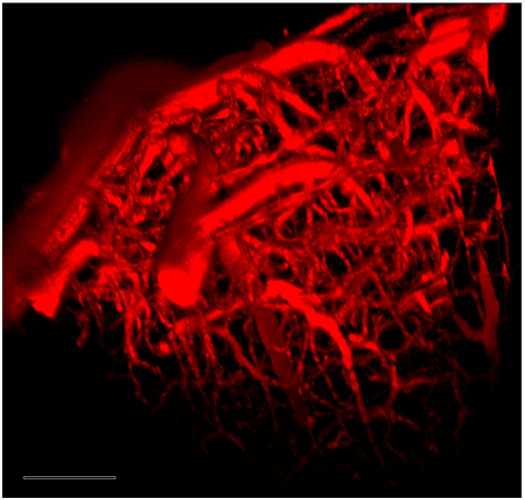
Image of the cerebral vascular system in the motor cortex of a mouse obtained by 3D two-photon microscopy with addition of Lem-PHEA dye (credit: B. van der Sanden and F. Appaix/Institut des Neurosciences de Grenoble)
A fluorescent dye that enables high-resolution (about 1 micron) 3D images of the cerebral vascular system has been synthesized by researchers at the Laboratoire de Chimie (CNRS) in France in collaboration with the Institut des Neurosciences (Université Joseph Fourier).
The “Lem-PHEA chromophore” dye fluoresces in the red-near infrared region biological transparency window using two-photon absorption and can pass through the skin. It features solubility in biological media, low cost, non-toxicity, and full elimination by the kidneys, suggesting that it may be suitable for in vivo imaging, the researchers say.
Injected into the blood vessels of a mouse, it has revealed details of the rodent’s vascular system with previously unattained precision, thanks to enhanced fluorescence compared to “conventional” dyes (such as Rhodamine-B and cyanine derivatives), and to conventional MRI medical imaging, which is limited to resolution of a few millimeters (1,000 times lower resolution), the researchers say.