Cells taken from the retina act as ‘ink’ in inkjet printer
December 18, 2013

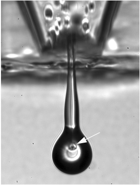
Close-up of retinal cells in a jet (credit: IOP Publising, Biofabrication)
UK researchers have used inkjet printing technology to successfully use ganglion cells and glial cells taken from the eye as “ink” in printing retinal patterns.
The breakthrough could lead to the production of artificial tissue grafts made from the variety of cells found in the human retina and may aid in the search to cure blindness.
The results are preliminary and provide proof-of-principle that an inkjet printer can be used to print the two types of intact cells from the retina of adult rats in patterns.
This is the first time the technology has been used successfully to print mature central nervous system cells and the results showed that printed cells remained healthy and retained their ability to survive and grow in culture.
The ability to arrange cells into highly defined patterns and structures has recently elevated the use of 3D printing in the biomedical sciences to create cell-based structures for use in regenerative medicine.
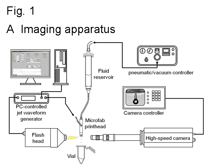
In their study, the researchers used a piezoelectric inkjet printer device that ejected the cells through a sub-millimeter diameter nozzle when a specific electrical pulse was applied. They also used high speed video technology to record the printing process with high resolution and optimised their procedures accordingly.
“In order for a fluid to print well from an inkjet print head, its properties, such as viscosity and surface tension, need to conform to a fairly narrow range of values. Adding cells to the fluid complicates its properties significantly,” commented Dr Wen-Kai Hsiao, another member of the team based at the Inkjet Research Centre in Cambridge.

Imaging apparatus (credit: IOP Publising, Biofabrication)
Once printed, a number of tests were performed on each type of cell to see how many of the cells survived the process and how it affected their ability to survive and grow.
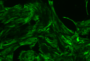
The cells derived from the retina of the rats were retinal ganglion cells, which transmit information from the eye to certain parts of the brain, and glial cells, which provide support and protection for neurons.
“We plan to extend this study to print other cells of the retina and to investigate if light-sensitive photoreceptors can be successfully printed using inkjet technology,” said Professor Keith Martin and Dr. Barbara Lorber, from the John van Geest Centre for Brain Repair, University of Cambridge.
“In addition, we would like to further develop our printing process to be suitable for commercial, multi-nozzle print heads.”
Making it available to patients
KurzweilAI asked Martin to comment on availability. “It is uncertain at this stage. This is currently a research tool, but an exciting one! At present this work provides proof of principle that retinal cells can be inkjet printed while retaining the ability to regrow processes essential to re-establishing functional neuronal networks. We aim to develop this technology to facilitate retinal repair in the context of degenerative, blinding eye diseases.
“There are similarities to what is being achieved with biological 3D printing in other fields, but this is the first time nerve cells from the mature adult central nervous system have been successfully inkjet printed.”
The work was funded by Fight for Sight, the van Geest Foundation and the EPSRC.
UPDATE 12/18/2013: wording changed in title and first paragraph to clarify printing process.
Abstract of Biofabrication paper
We have investigated whether inkjet printing technology can be extended to print cells of the adult rat central nervous system (CNS), retinal ganglion cells (RGC) and glia, and the effects on survival and growth of these cells in culture, which is an important step in the development of tissue grafts for regenerative medicine, and may aid in the cure of blindness. We observed that RGC and glia can be successfully printed using a piezoelectric printer. Whilst inkjet printing reduced the cell population due to sedimentation within the printing system, imaging of the printhead nozzle, which is the area where the cells experience the greatest shear stress and rate, confirmed that there was no evidence of destruction or even significant distortion of the cells during jet ejection and drop formation. Importantly, the viability of the cells was not affected by the printing process. When we cultured the same number of printed and non-printed RGC/glial cells, there was no significant difference in cell survival and RGC neurite outgrowth. In addition, use of a glial substrate significantly increased RGC neurite outgrowth, and this effect was retained when the cells had been printed. In conclusion, printing of RGC and glia using a piezoelectric printhead does not adversely affect viability and survival/growth of the cells in culture. Importantly, printed glial cells retain their growth-promoting properties when used as a substrate, opening new avenues for printed CNS grafts in regenerative medicine.
