Color-coding brain cells
December 23, 2014
University of Southampton neuroscientists have developed a method called “multicolor RGB tracking” to improve our understanding of how the brain works by color-marking individual brain cells in mice allows them to be tracked over space and time.*
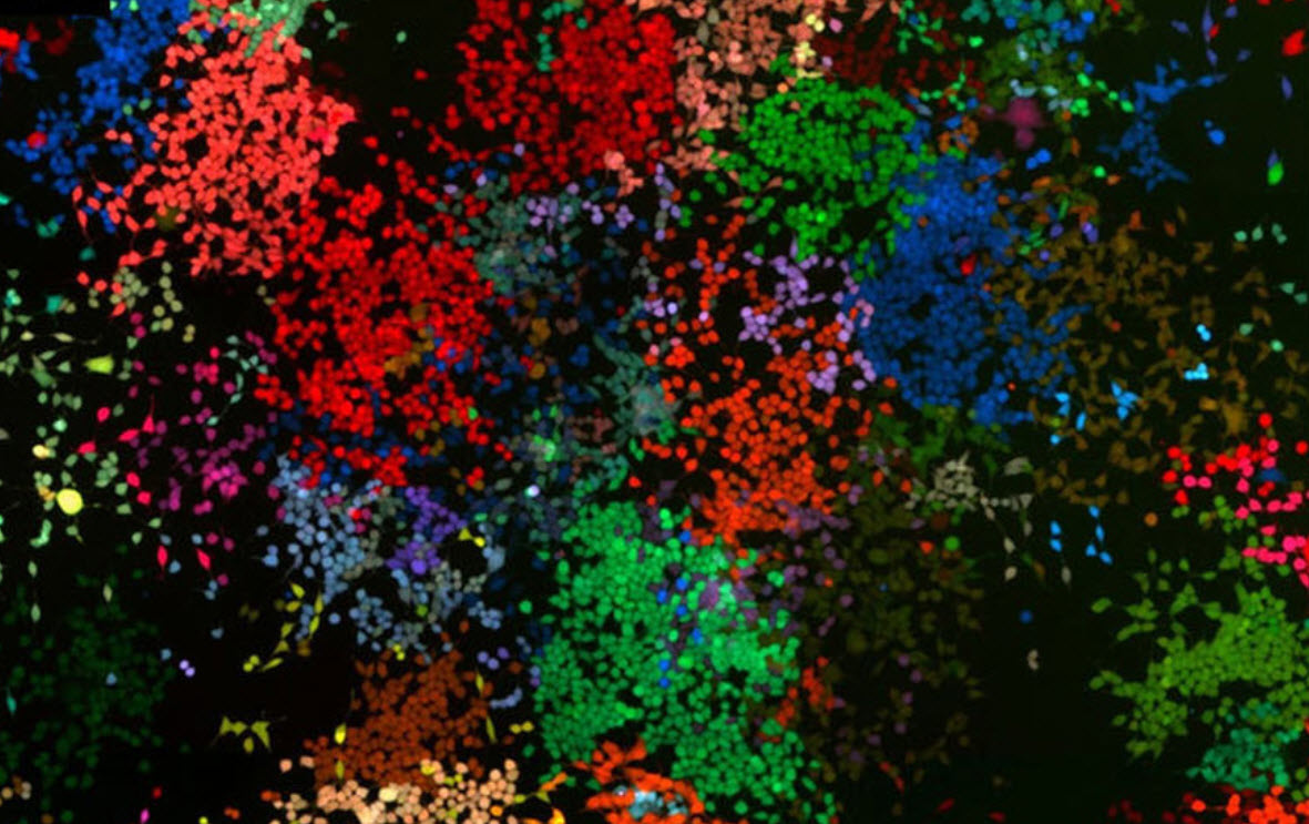
To mark a brain cell, they inject a solution that contains three viral vectors (delivery of genes by a virus) to create a fluorescent protein in each cell. Each cell then takes on a unique combination of the three colors (red, green and blue), acquiring a characteristic watermark. This approach allows researchers to color-code cells that would otherwise not be visible and distinguishable from each other.
Once the cell has been marked, the mark integrates into the DNA of the cell and will be expressed forever in that cell, as well as in any daughter cells.

RGB marking of human embryonic kidney cells grown in tissue culture (credit: Diego Gomez-Nicola et al./Scientific Reports)
According to Diego Gomez-Nicola, a Career Track Lecturer and MRC NIRG Fellow in the Centre for Biological Sciences at the University of Southampton, who led the multicolor RGB tracking research, “with this technique, we have proved the effective spatial and temporal tracking of neural cells, as well as the analysis of cell progeny. We predict that the use of multicolor RGB tracking will have an impact on how neuroscientists around the world design their experiments. It will allow them to answer questions they were unable to tackle before and contribute to the progress of understanding how our brain works.”
The researchers’ next step is to change the physiology or identity of certain cells by driving multiple genetic modification of genes of interest with the RGB vectors. In the same way they made cells express fluorescent proteins, researchers hope they can change the cell expression of specific genes of interest, which would serve as a basis for gene-therapy-based therapeutic approaches.
The research, which is published in the open-access journal Scientific Reports, was funded by the Medical Research Council (MRC), the EU and Wessex Medical Research and also involved researchers from the University of Hamburg, Germany.
* There are other brain-cell color schemes, notably the brainbow mouse, which has some disadvantages, including “limited cellular specificity of the fluorescent labeling, limited options for timing and spatial distribution of the labeling, restricted (immediate) availability for the broad scientific community, and the fact that even small modifications require time-consuming breeding programs,” the authors state in the paper.
Abstract of In-vivo RGB marking and multicolour single-cell tracking in the adult brain
In neuroscience it is a technical challenge to identify and follow the temporal and spatial distribution of cells as they differentiate. We hypothesised that RGB marking, the tagging of individual cells with unique hues resulting from simultaneous expression of the three basic colours red, green and blue, provides a convenient toolbox for the study of the CNS anatomy at the single-cell level. Using γ-retroviral and lentiviral vector sets we describe for the first time the in-vivo multicolour RGB marking of neurons in the adult brain. RGB marking also enabled us to track the spatial and temporal fate of neural stem cells in the adult brain. The application of different viral envelopes and promoters provided a useful approach to track the generation of neurons vs. glial cells at the neurogenic niche, allowing the identification of the prominent generation of new astrocytes to the striatum. Multicolour RGB marking could serve as a universal and reproducible method to study and manipulate the CNS at the single-cell level, in both health and disease.
