Combining magnetic sensing and imaging systems may improve brain diagnosis and imaging
July 27, 2012
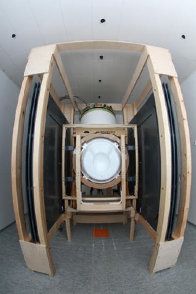
Innovative MEG-MRI device combines magnetoencephalography (MEG) and magnetic resonance imaging (MRI) technologies (credit: Aalto University)
The first system for mapping the human brain that combines whole-head magnetoencephalography (MEG) and magnetic resonance imaging (MRI) technology has been developed by a research team headed by Aalto University in Finland.
Merging these two technologies will produce unprecedented accuracy in locating and imaging brain electrical activity non-invasively, and should improve cancer diagnosis and the accuracy of brain mapping of patients, says professor Risto Ilmoniem.
Background
MEG measures the brain’s electrical activity, using highly sensitive superconducting quantum interference devices (SQUIDs) coupled to superconducting receiver (pickup) coils. MRI uses powerful magnets and radio frequency fields to image brain structure and blood flow.
When these two techniques are used separately, the image registration accuracy can be compromised because of the movement of the patient’s head, so the image it provides may not be accurate enough for precise brain surgery.
It has not been possible to combine MEG and MRI systems in the past because their magnetic fields interfered with one another. However, recently, MRI using SQUIDs has been demonstrated. This approach, called ultra-low-field (ULF) MRI, uses magnetic fields as low as 20 mT (microteslas) — 150,000 times weaker — for signal encoding, overcoming the magnetic interference problem.

MRI sensors, with MEG sensor mesh in the center (credit: Aalto University/Magnetic Resonance in Medicine)
Combining MEG and MRI technologies
Fusing these two technologies produces localization accuracy that was not possible with MRI or MEG alone. It will also allow for brain imaging of patients with metal implants, and will not scare children or make people feel claustrophobic.
The researchers were able to achieve MRI imaging quality comparable to 3T MRI machines and a commercial MEG device.
In the future, this development may also reduce costs because images can be obtained in one session rather than two, Ilmoniemi says.
The project is coordinated by Aalto University and includes 13 research groups in five countries. The research project is part of the European Commission Seventh Framework Program.