Custom 3-D printed ear models help surgeons carve new ears
October 21, 2015

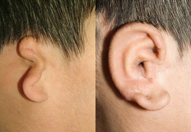
Children with under-formed or missing ears can undergo surgeries to fashion a new ear from rib cartilage, as shown in the above photo. But aspiring surgeons lack lifelike practice models. (credit: University of Washington)
A University of Washington (UW) otolaryngology resident and a bioengineering student have used 3-D printing to create a low-cost pediatric rib cartilage model that more closely resembles the feel of real cartilage, which is used in an operation called auricular reconstruction (ear replacement).
The innovation could make it possible for aspiring surgeons to become proficient in the sought-after but challenging procedure. And because the UW models are printed from a CT scan, they mimic an individual’s specific unique anatomy. That offers the opportunity for even an experienced surgeon to practice a particular tricky surgery ahead of time on a patient-specific rib model.
As part of the study, three experienced surgeons practiced carving, bending, and suturing the UW team’s silicone models, which were produced from a 3-D printed mold modeled from a CT scan of an 8-year-old patient. They compared their firmness, feel, and suturing quality to real rib cartilage, and to a more expensive material made out of dental impression material. They preferred the 3-D printed versions.

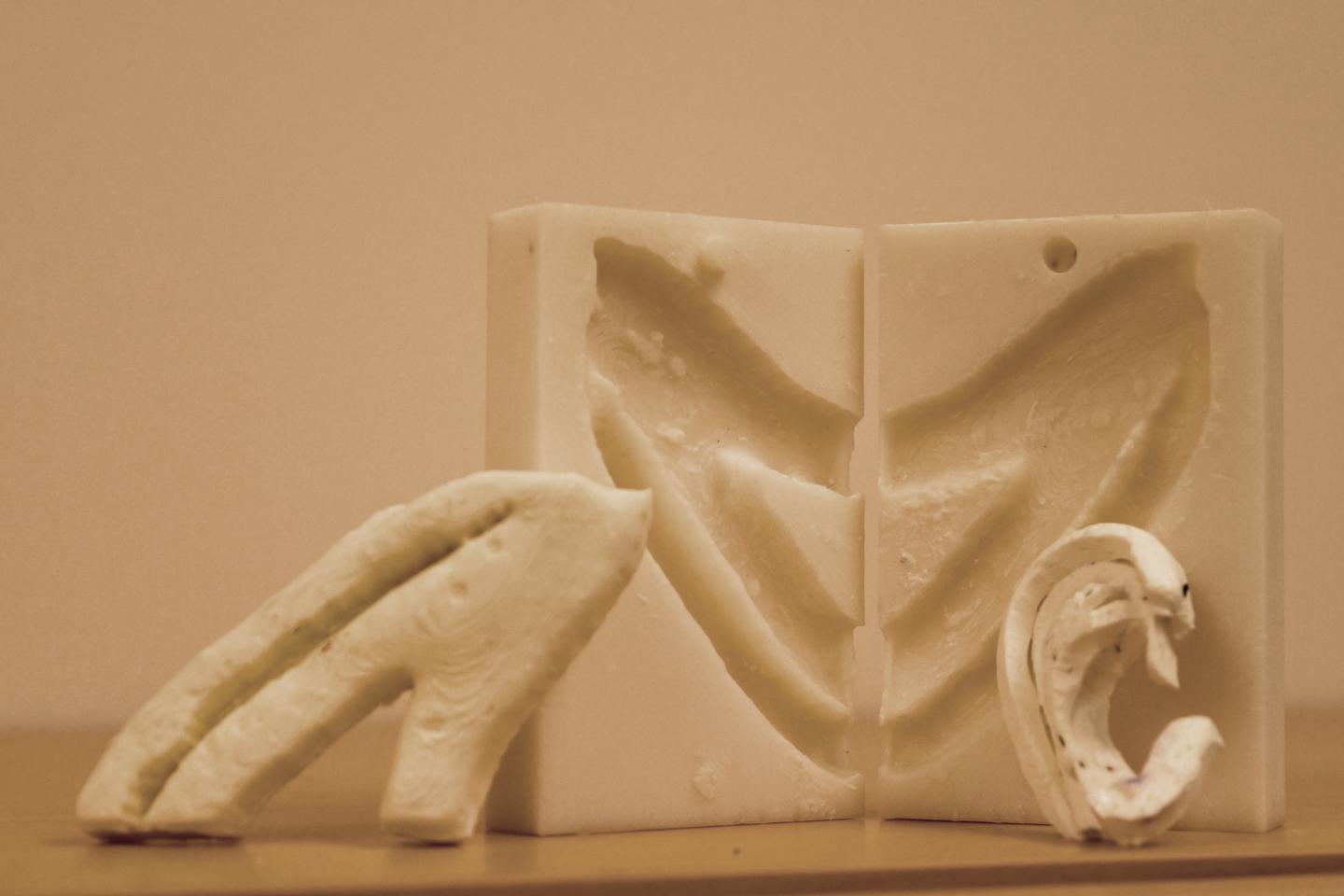
The UW team used a 3-D printer to create a negative mold of a patient’s ribs from a CT scan. Surgeons take pieces of those ribs and “carve” them into a new ear. (credit: University of Washington)
Co-author Sharon Newman, who graduated from the UW with a bioengineering degree in June, teamed up with lead author Angelique Berens, a UW School of Medicine otolaryngologist, while they both worked in the UW BioRobotics Lab under electrical engineering professor Blake Hannaford.
Newman figured out how to upload and process a CT scan through a series of free, open-source modeling and imaging programs, and ultimately use a 3-D printer to print a negative mold of a patient’s ribs.
Newman had previously tested different combinations of silicone, corn starch, mineral oil and glycerin to replicate human tissue that the lab’s surgical robot could manipulate. She poured them into the molds and let them cure to see which mixture most closely resembled rib cartilage.
The team’s next steps are to get the models into the hands of surgeons and surgeons-in-training, and hopefully to demonstrate that more lifelike practice models can elevate their skills and abilities.
“With one 3-D printed mold, you can make a billion of these models for next to nothing,” said Berens. “What this research shows is that we can move forward with one of these models and start using it.”
Long waiting list
Kathleen Sie, a UW Medicine professor of otolaryngology – head and neck surgery and director of the Childhood Communication Center at Seattle Children’s, said the lack of adequate training models makes it difficult for surgeons to become comfortable performing the delicate technical procedure.
There’s typically a six- to 12-month waiting list for children to have the procedure done at Seattle Children’s, she said.
“It’s a surgery that more people could do, but this is often the single biggest roadblock,” Sie said. “They’re hesitant to start because they’ve never carved an ear before.”
Their study results were presented at the American Academy of Otolaryngology — Head and Neck Surgery conference in Dallas.