Do autistic brains create more information at rest or do they have weaker connectivity — or both?
February 4, 2014

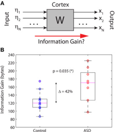
Information gain in the brain’s resting state. (A) Schematic black-box representation of cortical dynamics in the resting state. (B) The information gain is significantly increased by 42% in autistic relative to non-autistic children. (Credit: José L. Pérez Velázquez & Roberto F. Galán/Frontiers in Neuroinformatics)
New research from Case Western Reserve University and University of Toronto neuroscientists finds that the brains of autistic children generate more information at rest — a 42% increase on average.
The study offers a scientific explanation for the most typical characteristic of autism — withdrawal into one’s own inner world. The excess production of information may explain a child’s detachment from their environment.
Published in Frontiers in Neuroinformatics (open access), this study is a follow-up to the authors’ prior finding that brain connections are different in autistic children.
The authors quantified brain activity recorded with magnetoencephalography (MEG). “Our results suggest that autistic children are not interested in social interactions because their brains generate more information at rest, which we interpret as more introspection in line with early descriptions of the disorder,” said Roberto Fernández Galán, PhD, senior author and associate professor of neurosciences at Case Western Reserve School of Medicine.
The researchers also quantified interactions between brain regions (the brain’s functional connectivity), and determined the inputs to the brain in the resting state, allowing them to interpret the children’s introspection level.
This study provides quantitative support for the relatively new “Intense World Theory” of autism proposed by neuroscientists Henry and Kamila Markram of the Brain Mind Institute in Switzerland, which describes the disorder as the result of hyper-functioning neural circuitry, leading to a state of over-arousal.
More generally, the work of Galán and Pérez Velázquez is an initial step in the investigation of how information generation in the brain relates to cognitive/psychological traits and will begin to frame neurophysiological data into psychological aspects. The team now aims to apply a similar approach to patients with schizophrenia.
Galán is a scholar of The Mt. Sinai Health Care Foundation and former fellow of The Alfred P. Sloan Foundation.
Failure to eliminate links between neurons produces autistic-like mice

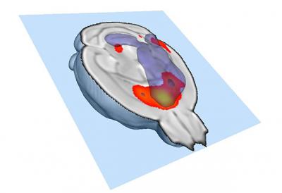
This image shows strongly connected brain regions. In mice with fewer microglia, the links between these regions are weaker. (Credit: Alessandro Gozzi, Istituto Italiano di Tecnologia)
In another study, just published in Nature Neuroscience, Italian neuroscientists suggested another explanation for autism: different parts of the brain don’t talk to each other very well, caused by microglia cells failing to trim connections between neurons.
The scientists used fMRI. Their findings indicate that by trimming surplus connections in the developing brain, microglia normally allow the remaining links to grow stronger, like high-speed fiber-optic cables carrying strong signals between brain regions.
But if microglia cells fail to do their job at that crucial stage of development, those brain regions are left with a weaker communication network, which in turn has lifelong effects on behavior, including decreased social behavior and increased repetitive behavior — hallmarks of autism.
The study involved scientists at the European Molecular Biology Laboratory (EMBL) in Monterotondo, Italy, and collaborators at the Istituto Italiano di Tecnologia (IIT), in Rovereto, and La Sapienza University in Rome.
Abstract of Frontiers in Neuroinformatics paper
Along with the study of brain activity evoked by external stimuli, an increased interest in the research of background, “noisy” brain activity is fast developing in current neuroscience. It is becoming apparent that this “resting-state” activity is a major factor determining other, more particular, responses to stimuli and hence it can be argued that background activity carries important information used by the nervous systems for adaptive behaviors. In this context, we investigated the generation of information in ongoing brain activity recorded with magnetoencephalography (MEG) in children with autism spectrum disorder (ASD) and non-autistic children. Using a stochastic dynamical model of brain dynamics, we were able to resolve not only the deterministic interactions between brain regions, i.e., the brain’s functional connectivity, but also the stochastic inputs to the brain in the resting state; an important component of large-scale neural dynamics that no other method can resolve to date. We then computed the Kullback-Leibler (KLD) divergence, also known as information gain or relative entropy, between the stochastic inputs and the brain activity at different locations (outputs) in children with ASD compared to controls. The divergence between the input noise and the brain’s ongoing activity extracted from our stochastic model was significantly higher in autistic relative to non-autistic children. This suggests that brains of subjects with autism create more information at rest. We propose that the excessive production of information in the absence of relevant sensory stimuli or attention to external cues underlies the cognitive differences between individuals with and without autism. We conclude that the information gain in the brain’s resting state provides quantitative evidence for perhaps the most typical characteristic in autism: withdrawal into one’s inner world.
“This is an exciting time to be studying microglia,” Gross concludes: “they’re turning out to be major players in how our brain gets wired up.”
Abstract of Nature Neuroscience paper
Microglia are phagocytic cells that infiltrate the brain during development and have a role in the elimination of synapses during brain maturation. Changes in microglial morphology and gene expression have been associated with neurodevelopmental disorders. However, it remains unknown whether these changes are a primary cause or a secondary consequence of neuronal deficits. Here we tested whether a primary deficit in microglia was sufficient to induce some autism-related behavioral and functional connectivity deficits. Mice lacking the chemokine receptor Cx3cr1 exhibit a transient reduction of microglia during the early postnatal period and a consequent deficit in synaptic pruning. We show that deficient synaptic pruning is associated with weak synaptic transmission, decreased functional brain connectivity, deficits in social interaction and increased repetitive-behavior phenotypes that have been previously associated with autism and other neurodevelopmental and neuropsychiatric disorders. These findings open the possibility that disruptions in microglia-mediated synaptic pruning could contribute to neurod