Dreamcatcher: scientists predict images seen in dreams
April 8, 2013

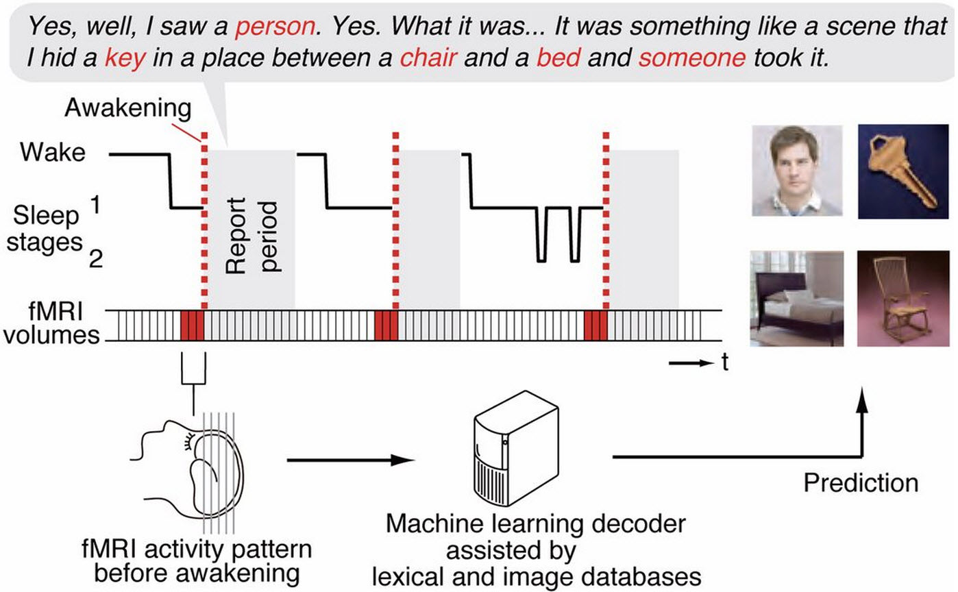
fMRI sleep experiments (credit: T. Horikawa et al./Science)
Japanese researchers have successfully predicted images seen in sleep based on MRI scans of brain activity.
As reported in Science, they recruited three volunteers to sleep in fMRI machines for 3-hour sessions over the course of 10 days while the researchers monitored each volunteer’s brain activity. They also used EEG to track the brain’s overall electrical activity.
The researchers woke subjects every six or seven minutes, asked them to report anything they had seen, then told them to go back to sleep. After gathering about 200 of these reports from each subject, the researchers extracted frequently repeated visual elements from the reports and grouped them into approximately 20 broad categories for each participant.

(A) Words describing visual objects or scenes (red) were mapped onto synsets of the WordNet tree. Synsets were grouped into base synsets (blue frames) located higher in the tree. (B) Visual reports (subject 2) are represented by visual content vectors, in which the presence/absence of the base synsets in the report at each awakening is indicated by white/black. (Credit: T. Horikawa et al./Science)
To train a computer program to associate broad categories of images seen in dreams with specific patterns of brain activity in the visual cortex, the researchers gathered photos on the Internet that corresponded to each category and recorded the three volunteers’ fMRI activity as they viewed the images.
The researchers then used the newly trained programs in a second round of dream-reporting by the same subjects, coupled with fMRI monitoring, and were able to predict what the people had seen with 60% accuracy.