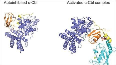
First 3D structural model of cancer-prevention molecule
January 27, 2012

The c-Cbl protein changes shape when switched on, marking a cell for destruction to prevent uncontrolled cell growth leading to cancer (credit: Hao Dou, et al./Nature Structural & Molecular Biology)
Cancer Research UK scientists have mapped the first 3D structure of c-Cbl, a key protein that protects against the development of cancer.
The team at Cancer Research UK’s Beatson Institute for Cancer Research, in Glasgow used X-ray analysis to map the structure of a protein called c-Cbl and showed that it changes shape when it is switched on.
c-Cbl controls cell growth , which when unregulated causes cells to divide excessively and can lead to cancer. The protein is defective in some leukemia patients. Discovering that c-Cbl can switch between two shapes may help scientists find ways to prevent faulty c-Cbl from triggering cancer.
“Understanding the structure of this protein is vital, because if the protein can’t be switched on, it is more likely to cause cancer,” said lead author, Dr. Danny Huang, at Cancer Research UK’s Beatson Institute in Glasgow. “So cracking the 3D structure is a step towards designing the cancer drugs of the future.”
The team showed that when it’s switched on, c-Cbl labels a cell receptor molecule for destruction. By labeling the receptor molecule for destruction, the cell growth signal is switched off at the right time. If c-Cbl cannot change to its active shape, it cannot label the receptor for destruction.
In healthy cells, the receptor amplifies a chain of cell signals, resulting in normal cell growth. But in cancer cells, these signals do not get switched off, leading to uncontrolled cell growth.
Ref.: Hao Dou, et al., Structural basis for autoinhibition and phosphorylation-dependent activation of c-Cbl, Nature Structural & Molecular Biology, 2012; [DOI:10.1038/nsmb.2231]