First map of core white-matter connections of human brain developed at USC
February 12, 2014

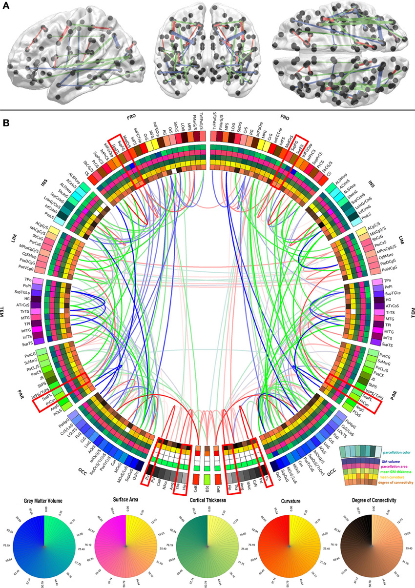
Graphical representation of the human-brain connectivity scaffold. Standard 3D graphs (A) and a connectogram (B) are used to visualize white-matter connections whose removal leads to significant changes in network integration and segregation. Only connections with this property are represented. (Credit: USC Institute for Neuroimaging and Informatics)
USC neuroscientists have systematically created the first map of the core white-matter “scaffold” (connections) of the human brain — the critical communications network that supports brain function.
Their work, published Feb. 11 in the open-access journal Frontiers in Human Neuroscience, has major implications for understanding brain injury and disease, the researchers say.
By detailing the connections that have the greatest influence over all other connections, the researchers offer a landmark first map of core white matter pathways and also show which connections may be most vulnerable to damage.
“We coined the term white matter ‘scaffold’ because this network defines the information architecture which supports brain function,” said senior author John Darrell Van Horn of the USC Institute for Neuroimaging and Informatics and the Laboratory of Neuro Imaging.
“While all connections in the brain have their importance, there are particular links which are the major players,” Van Horn said.
Using MRI data from a large sample of 110 individuals, lead author Andrei Irimia, also of the USC Institute for Neuroimaging and Informatics, and Van Horn systematically simulated the effects of damaging each white matter pathway.
Core pathways not limited to vulnerable gray-matter areas
They found that the most important areas of white and gray matter don’t always overlap. Gray matter is the outermost portion of the brain containing the neurons where information is processed and stored. Past research has identified the areas of gray matter that are disproportionately affected by injury.
But the current study shows that the most vulnerable white matter pathways — the core “scaffolding” — are not necessarily just the connections among the most vulnerable areas of gray matter, helping explain why seemingly small brain injuries may have such devastating effects.
“Sometimes people experience a head injury which seems severe but from which they are able to recover. On the other hand, some people have a seemingly small injury which has very serious clinical effects,” says Van Horn, associate professor of neurology at the Keck School of Medicine of USC. “This research helps us to better address clinical challenges such as traumatic brain injury and to determine what makes certain white matter pathways particularly vulnerable and important.”
“Such applications may be very useful to the task of determining how specific connectivity scaffold changes due either to gross pathology or to longitudinal white-matter atrophy can accumulate and ultimately produce appreciable neurological and cognitive deficits in TBI patients,” the researchers note in the paper.
The brain’s ‘social networks’
The researchers compare their brain imaging analysis to models used for understanding social networks. To get a sense of how the brain works, Irimia and Van Horn did not focus only on the most prominent gray matter nodes — which are akin to the individuals within a social network. Nor did they merely look at how connected those nodes are.
Rather, they also examined the strength of these white matter connections — which connections seemed to be particularly sensitive or to cause the greatest repercussions across the network when removed. Those connections that created the greatest changes form the network “scaffold.”
“Just as when you remove the internet connection to your computer you won’t get your email anymore, there are white matter pathways which result in large scale communication failures in the brain when damaged,” Van Horn said.
When white matter pathways are damaged, brain areas served by those connections may wither or have their functions taken over by other brain regions, the researchers explain.
Irimia and Van Horn’s research on core white matter connections is part of a worldwide scientific effort to map the 100 billion neurons and 1,000 trillion connections in the living human brain, led by the Human Connectome Project and the Laboratory of Neuro Imaging at USC.
Irimia notes that, “these new findings on the brain’s network scaffold help inform clinicians about the neurological impacts of brain diseases such as multiple sclerosis, Alzheimer’s disease, as well as major brain injury. Sports organizations, the military and the US government have considerable interest in understanding brain disorders, and our work contributes to that of other scientists in this exciting era for brain research.”
The research was supported by three NIH grants.
Abstract of Frontiers in Human Neuroscience paper
Brain connectivity loss due to traumatic brain injury, stroke or multiple sclerosis can have serious consequences on life quality and a measurable impact upon neural and cognitive function. Though brain network properties are known to be affected disproportionately by injuries to certain gray matter regions, the manner in which white matter (WM) insults affect such properties remains poorly understood. Here, network-theoretic analysis allows us to identify the existence of a macroscopic neural connectivity core in the adult human brain which is particularly sensitive to network lesioning. The systematic lesion analysis of brain connectivity matrices from diffusion neuroimaging over a large sample (N = 110) reveals that the global vulnerability of brain networks can be predicated upon the extent to which injuries disrupt this connectivity core, which is found to be quite distinct from the set of connections between rich club nodes in the brain. Thus, in addition to connectivity within the rich club, the brain as a network also contains a distinct core scaffold of network edges consisting of WM connections whose damage dramatically lowers the integrative properties of brain networks. This pattern of core WM fasciculi whose injury results in major alterations to overall network integrity presents new avenues for clinical outcome prediction following brain injury by relating lesion locations to connectivity core disruption and implications for recovery. The findings of this study contribute substantially to current understanding of the human WM connectome, its sensitivity to injury, and clarify a long-standing debate regarding the relative prominence of gray vs. WM regions in the context of brain structure and connectomic architecture.