Growing new brains with infrared light [exclusive]
May 24, 2013
Illustration of the “neuronal beacon” for guiding axon growth direction (credit: B. Black et al./Optics Letters)
University of Texas, Arlington, scientists have discovered a way to control the growth or repair of neurons and neuron circuits, using a non-invasive “neuronal beacon” (near-IR laser beam) — essentially rewiring brains, or even creating new ones.
This major discovery, just published today in Optics Letters, promises to enable several new applications, UT Arlington assistant professor of physics Samarendra Mohanty said in an exclusive interview with KurzweilAI:
- Building highly precise 3D neural circuits in-vitro as a model for future supercomputers using neuromorphic chips (or even using the neurons themselves in an artificially grown biological computer).
- Brain activity mapping, in combination with precision stimulation and imaging tools such as the fiber-optic, two-photon, optogenetic stimulator and label-free phase imaging developed by Mohanty.
- Repairing damaged neurons in the peripheral nervous system by rewiring around lesions (for patients with spinal-cord injuries, for example).
- Rewiring circuitry of the brain in the future, correcting for damaged or diseased neurons and neural circuits (the near-IR laser beam can penetrate deeply and non-invasively).
Guiding axon growth
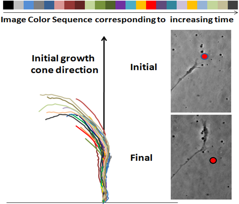
In a core discovery, Mohanty found that axon growth can be precisely controlled by shining a near-IR laser near the axon. This “neuronal beacon” process generates localized heat, causing the axon to change its growth direction in about 10 minutes. He found that axons can sense surprisingly low temperature rises (gradients) of less than 0.1 degrees C generated locally with a near-infrared laser.
Mohanty’s team performed optical-guidance experiments using cortical neurons isolated from embryonic 18-day rat embryos. A laser with beam power of 80 mW was reported, with a wavelength of 785 nm (700 to 1000 nm, and power of several orders of magnitude lower, also worked, but visible light caused damage to the axonal growth cone). The beam was placed ~5 µm away from the axons’ filopodia, asymmetrically positioned in the path of the advancing growth cone.
Creating neuronal networks

Laser-assisted navigation of rat cortical neurons. Left panel: time-lapse overlay of axonal shaft turning left, repelled by a laser beam. Right panel: image of a typical axon before and after the turn. The laser spot position is marked by the red circle. (Credit: B. Black et al./Optics Letters)
In the paper in Optics Letters, the authors say this neuronal-beacon method can be easily extended to form a neuronal network in-vitro by spatio-temporal control of the laser beam.
“This can be achieved by use of scanning laser beams or sculpting the laser spots by diffractive optical elements [like lenses], SLMs [spatial light modulators], and even standard light projectors.
“Being able to form in-vitro neuronal circuitry with high fidelity by the non-invasive photonic-guidance method described here will allow us to probe the functions of basic building blocks of the neuronal network.”
Nanosurgery
The neuronal circuitry can be augmented by ultrafast near-IR “laser scissors” for nanosurgery of undesired connections — silencing specific neurons or neuronal elements (axon, dendrite, spine) in the circuit. In combination with optical stimulation and imaging tools, this will enable all-optical testing of the computational nature of the neuronal circuit.”
Mohanty earlier demonstrated that the near-IR laser also allows for light-sensitization of neurons by transfection of opsin-encoding genes, and also for two-photon optogenetic stimulation and optical imaging. Mohanty believes rapid progress can be made in the all-optical control of neuronal circuit formation, and activity and mapping functional networks of the brain.
UPDATE May 27, 2013
According to Dr. Mohanty:
1. This discovery is primarily about a finding that central nervous system neurons (there may be others as well) are highly sensitive to a temperature gradient (<0.1 deg C). This is a fundamental discovery; it was not known earlier.
2. Since temperature is the cue (shown to be repulsive here, but can be attractive for other types of neurons), now there is a big possibility that very weakly focused light can do the guidance in large depths. In fact, pre-published data by the authors shows that this can be done non-invasively at depths of 4 mm, as compared to 100 µm done earlier (or fiber optically very near ~10 µm, see e.g., author’s prior work, S.K. Mohanty et al, J. Biomed. Opt. 13, 054049 (2008)). Further, any other method of temperature rise (not just lasers) can do it.
3. The current method to signal the growth cone can work at infrared laser power orders of magnitude lower than that used for prior force-based methods.
4. The authors employed this method for 360-degree guidance (results to be published) in primary neurons. No other method, optical or non-optical, has shown this. They have made complex circuits from normal cortical neurons that were earlier believed to be formed only in the case of genetic disorders.
5. The current discovery leads to 100% efficiency in retina-to-cortex guidance from mammals to fish, unlike the low efficiency in prior laser-based methods,.
6. The authors are already deploying their technique for in-vivo pre-clinical trials for spinal cord injury guidance.
7. Many researchers are biased towards guiding neurons by attractive means towards light and failing to look at the real repulsive effect of temperature rise. Irradiation of a primary neuron with intense light creates injury to a growth cone most of the time, but some researchers see some retraction or bending and misinterpret it as guidance.
For more information or to contact Dr. Samarendra Mohanty: http://www.uta.edu/faculty/smohanty/
A Google hangout is planned (information to be posted here) where authors will disclose the advancement of the technique with pathbreaking discoveries based on this technology.