High speed cancer-cell testing
April 3, 2013
Among a significant percentage of patients, the risk of metastasis of cancer is particularly expressed by the presence of an abnormal amount of protein HER2 on the surface of cancer cells.
A new in vitro system for identification of these proteins in diseased tissues. has been developed by EPFL and Institute of Pathology at the Lausanne University Hospital (CHUV) scientists. It is extremely fast, precise, inexpensive, and easy to use, the researchers say/
It also promises to enable healthcare providers to prescribe more effective customized treatment.
A diagnosis within minutes
The new diagnostic tool enables physicians to make an accurate assessment of the disease in a few minutes, in contrast to traditional methods, which require several hours.
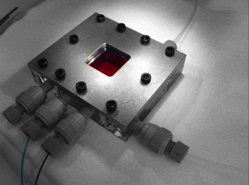
It comes in a chip made of silicon and glass, gridded by channels 100 microns in diameter. The principle is to make antibodies attach to the HER2 proteins of the cancerous tissue and detect them by fluorescence. The device can irrigate the entire sample evenly and for a set time.
“Currently, tissue samples must be bathed for a long time in a solution full of antibodies, so that each part of the tissue is thoroughly exposed,” says Martin Gijs, co-author of the publication and Director of the Laboratory of Microsystems (LMIS2). “But with this long period of immersion, the antibodies can disproportionately cling to targeted proteins or places where the proteins are not present, which gives ambiguous results. Frequently, complex and expensive genetic analysis (ISH) must then be additionally carried out.”
