How the brain organizes itself during development
August 4, 2011

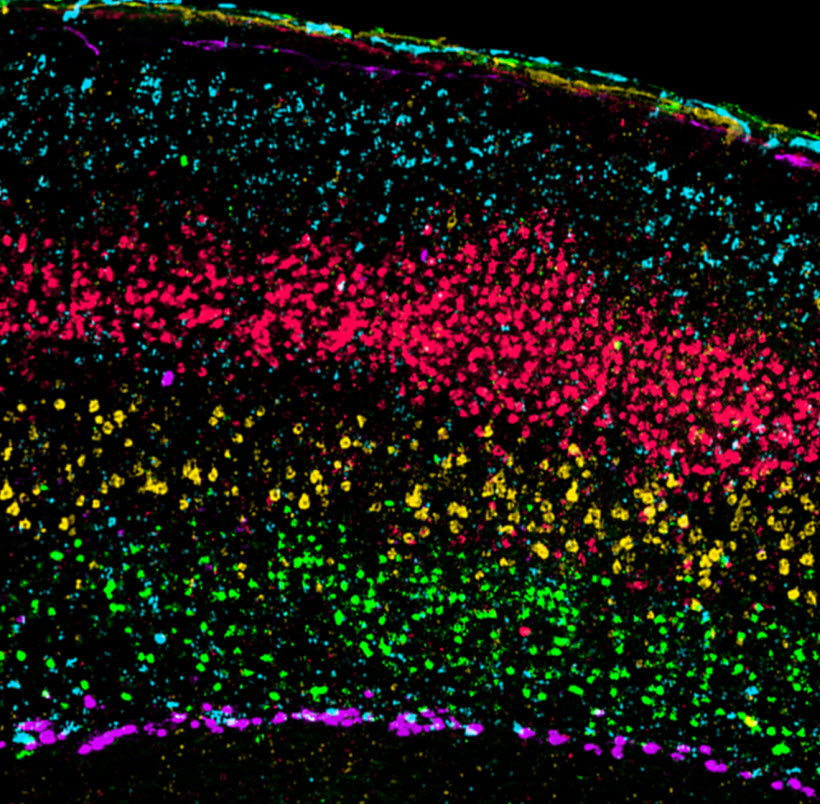
Strict segregation of layer-specific molecular markers in adult mouse somatosensory cortex. Pseudocolored overlay of serial brain sections labeled using fluorescent in situ hybridization for layer-specific cortical markers. (Credit: Maureen P. Boyle, et al./The Journal of Comparative Neurology)
Scientists at the Allen Institute for Brain Science have taken an important step in identifying how the brain organizes itself during development.
They call into question the current explanation of abnormal brain development in a well-studied strain of mouse known as reeler, named for its abnormal “reeling” gait, which has been integral in understanding how neurons migrate to their correct locations during brain development. The reeler cortex has been described for many years as being “inverted” compared to the normal neocortex.
However, the researchers have found that this abnormal layering is far more complex, more closely resembling a mirror-image inversion of normal cortical layering.
They used highly specific cellular markers, identified by mining the Allen Mouse Brain Atlas for genes with specific expression patterns. The Atlas is a genome-wide map of gene expression in the adult mouse brain. They employed the most precise molecular markers to date to identify features of cortical disorganization in the male reeler mouse that were unidentifiable using less-specific methods previously available.
The researchers observed unexpected cellular patterning that is difficult to explain by current models of neocortical development. The reeler mouse has a spontaneous mutation in a gene called Reelin that has been implicated in autism. Using in situ hybridization, a technique that allows for precise localization of specific genes, the researchers were able to follow developmental expression patterns through several stages of development to describe precise effects of Reelin deficiency in several brain areas during neurodevelopment.
The researchers identified, located, and tracked several specific cell types that are abnormally positioned in reeler mice.
Ref.: Maureen P. Boyle, et al., Cell-type-specific consequences of reelin deficiency in the mouse neocortex, hippocampus, and amygdala, The Journal of Comparative Neurology, 2011; 519 (11): 2061 [DOI: 10.1002/cne.22655]