Largest, sturdiest self-assembling DNA cages built
March 19, 2014

The five cage-shaped DNA polyhedra here have struts stabilizing their legs, and this innovation allowed a Wyss Institute team to build the largest and sturdiest DNA cages yet. The largest, a hexagonal prism (right), is one-tenth the size of an average bacterium. (Credit: Yonggang Ke/Harvard’s Wyss Institute)
Scientists at the Harvard’s Wyss Institute have built a set of self-assembling DNA cages that are up to one-tenth as wide as a bacterium. The structures are some of the largest and most complex structures ever constructed solely from DNA, they report in Science.
Moreover, the scientists visualized them using a DNA-based super-resolution microscopy method — and obtained the first sharp 3D optical images of intact synthetic DNA nanostructures in solution.
In the future, scientists could potentially coat the DNA cages to enclose their contents, packaging drugs for delivery to tissues. And, like a roomy closet, the cage could be modified with chemical hooks that could be used to hang other components such as proteins or gold nanoparticles. This could help scientists build a variety of technologies, including tiny power plants, miniscule factories that produce specialty chemicals, or high-sensitivity photonic sensors that diagnose disease by detecting molecules produced by abnormal tissue.
“I see exciting possibilities for this technology,” said Peng Yin, Ph.D., a Core Faculty member at the Wyss Institute and Assistant Professor of Systems Biology at Harvard Medical School, and senior author of the paper.
Building with DNA

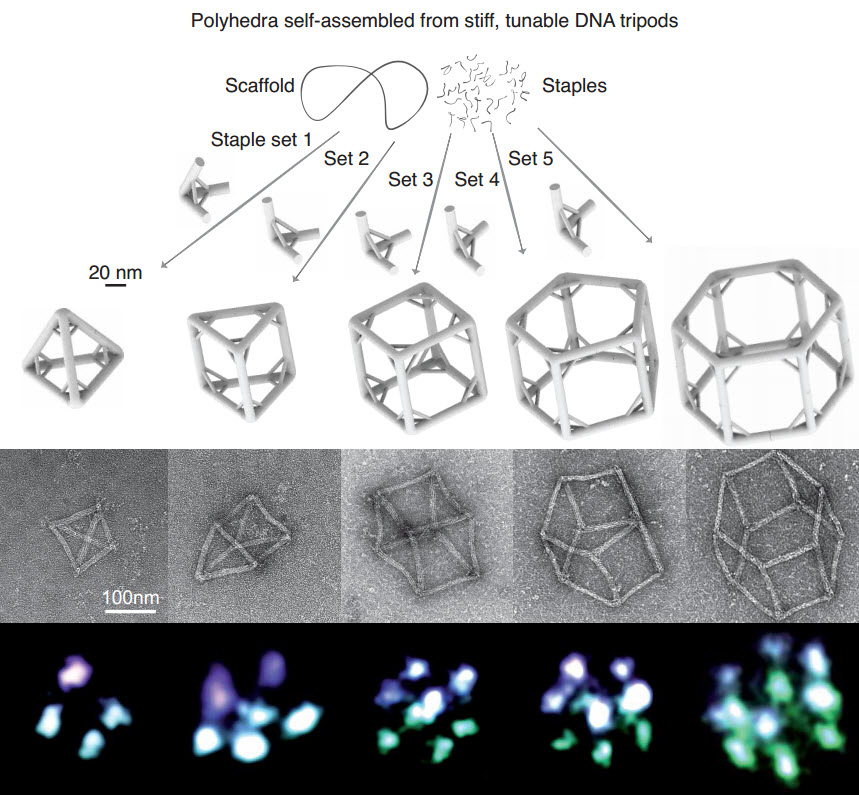
Wireframe polyhedra assembled from tunable DNA-origami tripods. Top: schematics showing the assembly process of tripod monomers and the polyhedra; middle: TEM images of polyhedra; bottom: super-resolution fluorescence images of polyhedra. (Credit: Ryosuke Iinuma et al./Science)
Scientists in the emerging field of DNA nanotechnology are exploring ways to use DNA to build tiny structures for a variety of applications. These structures are programmable, in that scientists can specify the sequence of letters, or bases, in the DNA, and those sequences then determine the structure it creates.
So far, most researchers in the field have used a method called DNA origami, in which short strands of DNA staple two or three separate segments of a much longer strand together, causing that strand to fold into a precise shape. DNA origami was pioneered in part by Wyss Institute Core Faculty member William Shih, Ph.D., who is also an Associate Professor in the Department of Biological Chemistry and Molecular Pharmacology at Harvard Medical School and the Department of Cancer Biology at the Dana-Farber Cancer Institute.
Yin’s team has built different types of DNA structures, including a modular set of parts called single-stranded DNA tiles or DNA bricks. Like LEGO bricks, these parts can be added or removed independently. Unlike LEGO bricks, they spontaneously self-assemble.
But for some applications, scientists might need to build much larger DNA structures than anyone has built so far. So, to add to their toolkit, Yin’s team sought much larger building blocks to match.
Engineering challenges

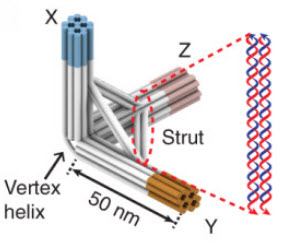
Design diagram of a DNA-origami tripod showing added strut for stability. Cylinders represent DNA double helices. (Credit: Ryosuke Iinuma et al./Science)
Yin and his colleagues first used DNA origami to create extra-large building blocks the shape of a photographer’s tripod. The plan was to engineer those tripod legs to attach end-to-end to form polyhedra — objects with many flat faces that are themselves triangles, rectangles, or other polygons.
But when Yin and associates built bigger tripods and tried to assemble them into polyhedra, the large tripods’ legs would splay and wobble, which kept them from making polyhedra at all.
The researchers got around that problem by building in a horizontal strut to stabilize each pair of legs, just as a furniture maker would use a piece of wood to bridge legs of a wobbly chair.
To glue the tripod legs together end-to-end, they took advantage of the fact that matching DNA strands pair up and adhere to each other. They left a tag of DNA hanging off a tripod leg, and a matching tag on the leg of a different tripod that they wanted it to pair with.
The team programmed DNA to fold into sturdy tripods 60 times larger than previous DNA tripod-like building blocks and 400 times larger than DNA bricks. Those tripods then self-assembled into a specific type of three-dimensional polyhedron — all in a single test tube.
By adjusting the length of the strut, they built tripods that ranged from upright to splay-legged. More upright tripods formed polyhedra with fewer faces and sharper angles, such as a tetrahedron, which has four triangular faces. More splay-legged tripods formed polyhedra with more faces, such as a hexagonal prism, which is shaped like a wheel of cheese and has eight faces, including its top and bottom.
In all, they created five polyhedra: a tetrahedron, a triangular prism, a cube, a pentagonal prism, and a hexagonal prism.
Ultrasharp snapshots
After building the cages, the scientists visualized them using a DNA-based microscopy method Jungmann had helped developed called DNA-PAINT. In DNA-PAINT, short strands of modified DNA cause points on a structure to blink, and data from the blinking images reveal structures too small to be seen with a conventional light microscope. DNA-PAINT produced ultrasharp snapshots of the researchers’ DNA cages — the first 3D snapshots ever of single DNA structures in their native, watery environment.
“Bioengineers interested in advancing the field of nanotechnology need to devise manufacturing methods that build sturdy components in a highly robust manner, and develop self-assembly methods that enable formation of nanoscale devices with defined structures and functions,” said Wyss Institute Founding Director Don Ingber, M.D., Ph.D. “Peng’s DNA cages and his methods for visualizing the process in solution represent major advances along this path.”
This work was funded by the Office of Naval Research, the Army Research Office, the National Institutes of Health, the National Science Foundation, the JSR Corporation, and the Wyss Institute.
Abstract of Science paper
DNA self-assembly has produced diverse synthetic three-dimensional polyhedra. These structures typically have a molecular weight no greater than 5 megadaltons (MD). We report a simple, general strategy for one-step self-assembly of wireframe DNA polyhedra that are more massive than most previous structures. A stiff three-arm-junction DNA origami tile motif with precisely controlled angles and arm lengths was used for hierarchical assembly of polyhedra. We experimentally constructed a tetrahedron (20 MD), a triangular prism (30 MD), a cube (40 MD), a pentagonal prism (50 MD), and a hexagonal prism (60 MD) with edge widths of 100 nanometers. The structures were visualized by transmission electron microscopy and by three-dimensional DNA-PAINT super-resolution fluorescent microscopy of single molecules in solution.