Milestone reached in building replacement kidneys in the lab
September 10, 2014

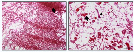
Blood-vessel clotting (left) after a few hours with previous kidney-implant method not found with new method (right) (credit: In Kap Ko et al./Technology)
Regenerative medicine researchers at Wake Forest Baptist Medical Center in North Carolina have developed what they say is the most successful method to date to keep blood vessels in new human-sized pig kidney organs open and flowing with blood — a major challenge in the quest to build replacement kidneys in the lab.
The work is reported in the journal Technology.
“According to the Urologic Diseases Information Clearinghouse, more than 870,000 people in the U.S. were undergoing treatment for ESRD in 2009. Current treatments are limited to life-long dialysis … which cannot replace many of the other functions of the kidneys, resulting in complications such as anemia, malnutrition, and eventually requiring [the other treatment option] kidney transplantation.” — Kap Ko et al./Technology
“Until now, lab-built kidneys have been rodent-sized and have functioned for only one or two hours after transplantation because blood clots developed,” said Anthony Atala, M.D., director and professor at the Wake Forest Institute for Regenerative Medicine and a senior author on the study.
“In our proof-of-concept study, the [blood] vessels in a human-sized pig kidney remained open during a four-hour testing period. We are now conducting a longer-term study to determine how long flow can be maintained.”
If proven successful, the new method to more effectively coat the blood vessels with endothelial cells (which line the interior surface of blood vessels) could potentially be applied to other complex organs that scientists are working to engineer, including the liver and pancreas, the researchers say.
How to create a replacement kidney that survives
The research is part of a long-term project to use pig kidneys to make scaffolds (support structures) that could potentially be used to build replacement kidneys for human patients who have end-stage renal disease. The main problem is dealing with blood clotting.
The current process fails within a few hours:
1. To avoid immune rejection, scientists currently first remove all animal cells (proteins and DNA) from the organ — leaving only the organ structure or “skeleton”(this is known as “decellularization.” A patient’s own cells are then be placed in the scaffold (“recellularization”), making an organ that the patient (theoretically) would not reject.
2. The cell removal process leaves behind an intact network of blood vessels that can potentially supply the new organ with oxygen. However, scientists working to repopulate kidney scaffolds with cells have had problems coating the vessels, so severe clotting has generally occurred within a few hours after transplantation.
The new Wake Forest Baptist approach looks more promising:

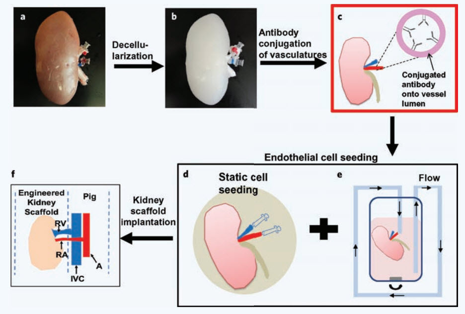
Schematic diagram of decellularization (b) of the pig kidney scaffold, adding antibodies (c) to help seed an endothelial blood-vessel layer (d, e), and in vivo implantation of the recellularized kidney (f) (credit: In Kap Ko et al./Technology)
1. First, they evaluated four different methods of introducing new cells into the main vessels of the kidney scaffold. They found that a combination of infusing cells with a syringe followed by a period of pumping cells through the vessels at increasing flow rates was most effective.
2. Next, the research team coated the scaffold’s blood vessels with an antibody designed to make them more “sticky” and to bind endothelial cells. Laboratory and imaging studies — as well as tests of blood flow in the lab — showed that cell coverage of the vessels was sufficient to support blood flow through the entire kidney scaffold.
3. The final test of the dual-approach was implanting the scaffolds in pigs weighing 90 to 110 pounds. During a four-hour testing period, the vessels remained open.
“Our cell-seeding method, combined with the antibody, improves the attachment of cells to the vessel wall and prevents the cells from being detached when blood flow is initiated,” said In Kap Ko, Ph.D., lead author and instructor in regenerative medicine at Wake Forest Baptist.
A step closer to replacing kidneys
However, a long-term examination is necessary to sufficiently conclude that blood clotting is prevented when endothelial cells are attached to the vessels, the scientists said.
They also noted that if the new method is proven successful in the long-term, the research brings them an important step closer to the day when replacement kidneys can be built in the lab.
“The results are a promising indicator that it is possible to produce a fully functional vascular system that can deliver nutrients and oxygen to engineered kidneys, as well as other engineered organs,” said Ko.
Why use pig kidneys as scaffolds for human patients? Mainly because the organs are similar in size to human kidneys and because pig heart valves — removed of cells — have safety been used in patients for more than three decades.
This study was supported, in part, by the Telemedicine and Advanced Technology Research Center at the U.S. Army Medical Research and Materiel Command.
Abstract of Technology paper
Decellularization of whole organs, such as the kidney hold great promise in addressing donor shortage for transplantation. However, successful implantation of engineered whole kidney constructs has been challenged by the inability to maintain endothelial cell coverage of the vasculature matrix, resulting in excessive blood clots, loss of vascular patency, and cell death within the construct. In this study, we describe an endothelial cell seeding approach that permits effective coating of the vascular matrix of the decellularized porcine kidney scaffold using a combination of static and ramping perfusion cell seeding. Furthermore, conjugation of CD31 antibodies to the vascular matrix improved endothelial cell retention on the vasculatures, which enhanced vascular patency of the implanted scaffold. These results demonstrate that our endothelial cell seeding method combined with antibody conjugation improves endothelial cell attachment and retention leading to vascular patency of tissue-engineered whole kidney in vivo.