Nanotools for neuroscience and brain activity mapping
March 25, 2013
“Neuroscience — one of the greatest challenges facing science and engineering — is at a crossroads. …There exist few general theories or principles that explain brain function [due partly to] limitations in current methodologies,” say neuroscientists in a new ACS Nano open-access paper, “Nanotools for Neuroscience and Brain Activity Mapping.”
Traditional neurophysiological approaches record the activities of one neuron or a few neurons at a time. Neurochemical approaches focus on single neurotransmitters. Yet, there is an increasing realization that neural circuits operate at emergent levels, where the interactions between hundreds or thousands of neurons, utilizing multiple chemical transmitters, generate functional states.
Entirely new tools will ultimately be required both to study neurons and neural circuits with minimal perturbation and to study the human brain.
The Brain Activity Map (BAM) project has been proposed* to develop such tools. It has three goals:
- Build neuroscience tools capable of:measuring the activity of large sets of neurons in complex brain circuits, with simultaneous measurement and manipulation of activity of thousands or even millions of neurons.
- Computationally analyze and model these brain circuits.
- Test these models by manipulating the activities of chosen sets of neurons in these brain circuits.
With access to 1 million neurons, scientists will be able to evaluate the function of the entire brain of the zebrafish or several areas from the cerebral cortex of the mouse.
In parallel, the neuroscientists envision developing nanoscale neural probes that can locally acquire, process, and store accumulated data. Networks of “intelligent” nanosystems would be capable of providing specific responses to externally applied signals, or to their own readings of brain activity.
Brain activity mapping tools
Nanofabricated planar electrode array for high-density neuronal voltage recording (credit: Du et al.)
Extracellular microelectrodes can resolve single-neuron firing in vivo without penetrating the cell.
To achieve this, the detection range is typically limited to neurons whose cell bodies are closer than ∼50 microns from the microelectrode surface.
Researchers can now deploy simultaneously nearly 1000 measurement sites distributed across several cortical areas of the same animal, allowing the firing patterns of hundreds of neurons to be monitored in parallel.

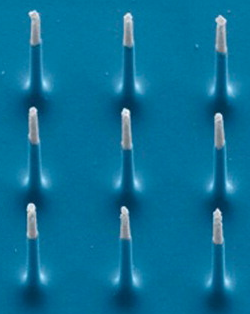
Silicon nanoneedles that constitute the active region of a 3D nanoelectrode array (credit: Nature Publishing Group)
This allows for monitoring multiple anatomical areas or sub-regions in tandem, and be scaled up in the future to sample arbitrarily complex networks.
“Electrodes can access deep brain structures that are challenging to reach with optical methods, they do not require labeling cells with a dye,and they offer higher sampling speed than voltage or calcium indicators,” the authors point out.
“Furthermore, the manufacturing processes used in voltage-sensing electrode development can translate to other modes of interrogating neuronal activity, such as chemical sensors.:
Nanoscale needle electrodes can provide high-fidelity electrophysiological interfaces and can perform intracellular recording and stimulation of neurons in a highly scalable fashion.

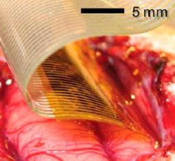
Flexible, high-density active electrode arrays in cat brain (credit: Nature Publishing Group)
Flexible and active electronics offer another potential option for interfaces to neural circuits and the brain. Significant progress has been made in the areas of ultrathin, flexible, light, biocompatible circuits.
Hundreds of contacts to the brain can be made on double-sided active semiconductor nanomembrane electronics transferred onto silk or other flexible substrates.
New Imaging Tools
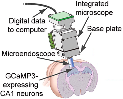
High-speed, miniaturized epi-fluorescence microscopes have been fabricated from mass-producible optical parts, such as light-emitting diodes (LEDs) and nanofabricated semiconductor sensors.
An adult mouse can readily bear such a microscope on the head during active behavior, which routinely allows imaging of calcium dynamics in >1000 neurons per mouse.
 Optogenetics allows for cellular genetic identity by “phototagging.” Optogenetic tools are themselves nanoscale devices that can be engineered for new classes of brain activity mapping function, building on molecular structure function relationships.
The BAM Project may bring us closer to answering ultimate questions about how we think and make decisions, which involve the coordinated activity in large numbers of neurons widely distributed throughout the human brain.
* Last month the New York Times revealed that the Obama Administration intends to seek billions of dollars from Congress for a Brain Activity Map (BAM) project, partly based on the paper “The Brain Activity Map Project and the Challenge of Functional Connectomics,” published on Neuron in June 2012. A more detailed open access version of the paper, with intriguing suggestions of fleets of nanobots swarming the living brain and recording the details of neural activity, is available online.
“The Brain Activity Map”(Science March 2013) noted that BAM could put neuroscientists in a position to understand how the brain produces perception, action, memories, thoughts, and consciousness. Within 5 years, it should be possible to monitor and/or to control tens of thousands of neurons, and by year 10 that number will increase at least 10-fold. By year 15, observing 1 million neurons with markedly reduced invasiveness should be possible.
