New catheter lets doctors see inside arteries for first time
October 5, 2016

Image-guided catheter with a camera the size of a grain of salt (credit: Avinger)
A new safer catheter design that allows cardiologists to see inside arteries for the first time and remove plaque from only diseased tissue has been used by interventional cardiologists at UC San Diego Health.
The new image-guided device, Avinger’s Pantheris, allows doctors to see and remove plaque simultaneously during an atherectomy — a minimally invasive procedure that involves cutting plaque away from the artery and clearing it out to restore blood flow.

Left: OCT imaging fiber on cutter allows for seeing the layers and disease. Center: the torque shaft, cutter window, and apposition balloon prove the direction and precision to avoid disruption of layers. Right: the cutter and nose-cone allow for capturing and removing only diseased tissue. (credit: Avinger)
The new technology treats patients suffering from the painful symptoms of peripheral artery disease (PAD), a condition caused by a build-up of plaque that blocks blood flow in the arteries of the legs and feet, preventing oxygen-rich blood from reaching the extremities. Patients with PAD frequently develop life threatening complications, including heart attack and stroke; in some severe cases, patients may even face amputation.
PAD affects nearly 20 million adults in the United States and more than 200 million globally.
“Peripheral artery disease greatly impacts quality of life, with patients experiencing cramping, numbness and discoloration of their extremities,” said Mitul Patel, MD, cardiologist at UC San Diego Health. “This new device is a significant step forward for the treatment of PAD with a more efficient approach for plaque removal and less radiation exposure to the doctor and patient.”

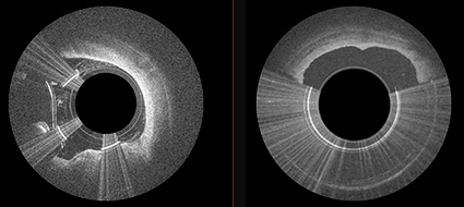
Pantheris allows interventional cardiologists to see the layers and disease (left) and remove just the disease (right) (credit: Avinger)
X-ray technology was previously used during similar procedures, but those images are not as clear and do not allow for visualization inside the blood vessel. The new catheter, with a fiber optic camera the size of a grain of salt on the tip, is fed through a small incision in the groin that does not require full anesthesia. Once inside, the interventional cardiologist is able to see exactly what needs to be removed without damaging the artery wall, which can cause further narrowing.
Pantheris was approved by the FDA in March 2016. So far, cardiologists at UC San Diego Health have used the new catheter on 10 patients undergoing an atherectomy procedure, with successful results.
Avinger | PML0290-D Pantheris Animation
Abstract of Utility of Image-Guided Atherectomy for Optimal Treatment of Ambiguous Lesions by Angiography
Peripheral endovascular interventions have been limited by multiple shortcomings, including difficulty in assessment of the 3-dimensional nature of obstructive plaque within the vasculature with only contrast angiography and 2-dimensional fluoroscopy. Herein, we present a case of pseudodissection seen angiographically post CTO crossing, which was accurately assessed as eccentric plaque using OCT imaging and treated using an OCT-guided directional atherectomy device, preventing bail-out stenting.