New diagnostic chip can generate single-cell molecular ‘fingerprints’ for brain tumors
August 4, 2010

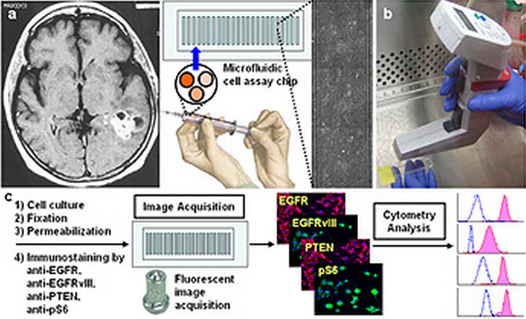
Conceptual summary of the microfluidic image cytometry technology: (a) Dissociated glioblastoma cells obtained from a patient are introduced into a microfluidic cell array chip for multi-parameter analysis by this technology. (b) A semi-automated pipette executes cell seeding/culture and ICC. (c) The ICC-treated samples in the chips are mounted on a fluorescent microscope for image acquisition followed by analysis using an image cytometry program (i.e., Metamorph, Molecular Devices Inc.) to quantify the expression levels of signaling proteins with single-cell resolution.
UCLA researchers have developed a cancer diagnostic chip that generates single-cell “molecular fingerprints” for a small quantity of pathology samples, including brain tumor tissues, combining the advantages of microfluidics and microscopy-based cell imaging.
The new microfluidic image cytometry (MIC) platform is capable of making molecular measurements on small tumor samples provided by tumor resection and biopsy using as few as 1,000 to 3,000 cells, according to the researchers.
“The promise and attractiveness of this approach is the small amount of tissue needed for analysis in the face of increasing numbers of prognostic and predictive markers,” said Dr. William Yong, a Jonsson Cancer Center physician-scientist who led the pathology aspects of the research.
The researchers will next apply the new platform to larger cohorts of cancer patient samples and integrate the diagnostic approach into clinical trials of molecular therapies.
This study was funded by the National Cancer Institute/ National Institutes of Health (NCI/NIH) and the National Institute of Neurological Disorders and Stroke (NINDS).
More info: UCLA news