New nuclear magnetic resonance technique offers ‘molecular window’ for live disease diagnosis
May 3, 2017

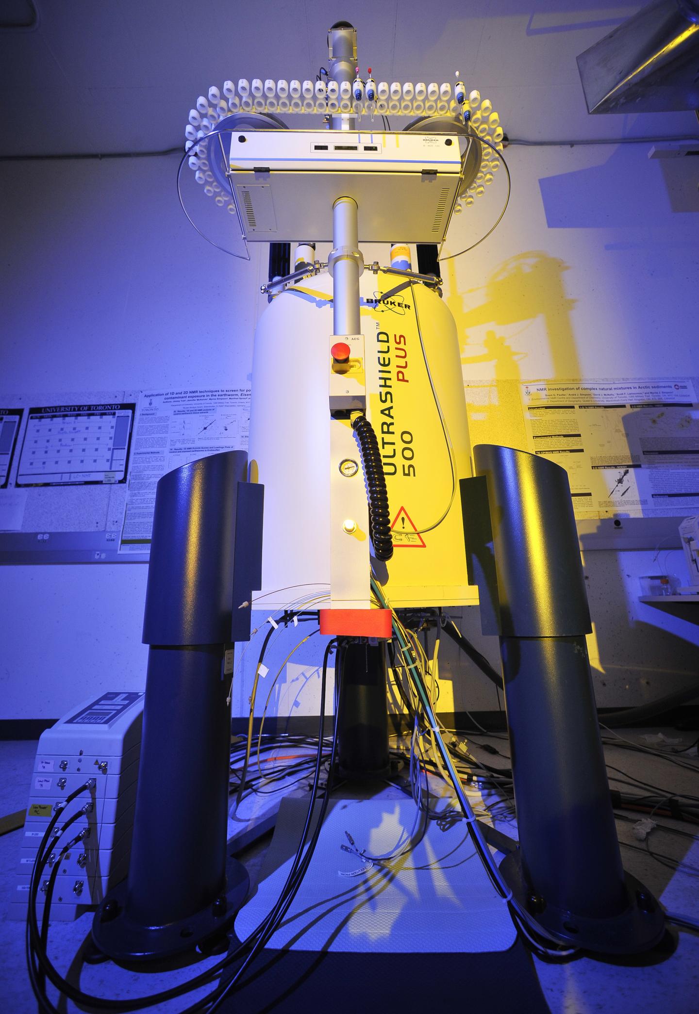
New nuclear magnetic resonance (NMR) system for molecular diagnosis (credit: University of Toronto Scarborough)
University of Toronto Scarborough researchers have developed a new “molecular window” technology based on nuclear magnetic resonance (NMR) that can look inside a living system to get a high-resolution profile of which specific molecules are present, and extract a full metabolic profile.
“Getting a sense of which molecules are in a tissue sample is important if you want to know if it’s cancerous, or if you want to know if certain environmental contaminants are harming cells inside the body,” says Professor Andre Simpson, who led research in developing the new technique.*

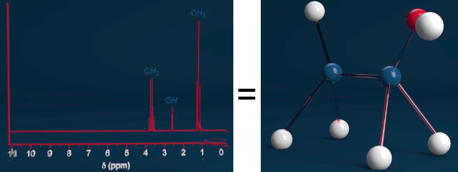
An NMR spectrometer generates a powerful magnetic field that causes atomic nuclei to absorb and re-emit energy in distinct patterns, revealing a unique molecular signature — in this example: the chemical ethanol. (credit: adapted from the Bruker BioSpin “How NMR Works” video at www.theresonance.com/nmr-know-how)
Simpson says there’s great medical potential for this new technique, since it can be adapted to work on existing magnetic resonance imaging (MRI) systems found in hospitals. “It could have implications for disease diagnosis and a deeper understanding of how important biological processes work,” by targeting specific biomarker molecules that are unique to specific diseased tissue.
The new approach could detect these signatures without resorting to surgery and could determine, for example, whether a growth is cancerous or benign directly from the MRI alone.
The technique could also provide highly detailed information on how the brain works, revealing the actual chemicals involved in a particular response. “It could mark an important step in unraveling the biochemistry of the brain,” says Simpson.
Overcoming magnetic distortion
Until now, traditional NMR techniques haven’t been able to provide high-resolution profiles of living organisms because of magnetic distortions from the tissue itself. Simpson and his team were able to overcome this problem by creating tiny communication channels based on “long-range dipole interactions” between molecules.
The next step for the research is to test it on human tissue samples, says Simpson. Since the technique detects all cellular metabolites (substances such as glucose) equally, there’s also potential for non-targeted discovery.
“Since you can see metabolites in a sample that you weren’t able to see before, you can now identify molecules that may indicate there’s a problem,” he explains. “You can then determine whether you need further testing or surgery. So the potential for this technique is truly exciting.”
The research results are published in the journal Angewandte Chemie.
* Simpson has been working on perfecting the technique for more than three years with colleagues at Bruker BioSpin, a scientific instruments company that specializes in developing NMR technology. The technique, called “in-phase intermolecular single quantum coherence” (IP-iSQC), is based on some unexpected scientific concepts that were discovered in 1995, which at the time were described as impossible and “crazed” by many researchers. The technique developed by Simpson and his team builds upon these early discoveries. The work was supported by Mark Krembil of the Krembil Foundation and the Natural Sciences Engineering Research Council of Canada (NSERC).
Abstract of In-Phase Ultra High-Resolution In Vivo NMR
Although current NMR techniques allow organisms to be studied in vivo, magnetic susceptibility distortions, which arise from inhomogeneous distributions of chemical moieties, prevent the acquisition of high-resolution NMR spectra. Intermolecular single quantum coherence (iSQC) is a technique that breaks the sample’s spatial isotropy to form long range dipolar couplings, which can be exploited to extract chemical shift information free of perturbations. While this approach holds vast potential, present practical limitations include radiation damping, relaxation losses, and non-phase sensitive data. Herein, these drawbacks are addressed, and a new technique termed in-phase iSQC (IP-iSQC) is introduced. When applied to a living system, high-resolution NMR spectra, nearly identical to a buffer extract, are obtained. The ability to look inside an organism and extract a high-resolution metabolic profile is profound and should find applications in fields in which metabolism or in vivo processes are of interest.