Organic transistors for brain mapping
April 15, 2013

This electrocorticography (ECoG) sensor array with transistors can now obtain direct, high-quality amplification and brain-signal recording (credit: Department of Bioelectronics, Ecole des Mines)
To improve brain mapping, a group of French scientists have produced the world’s first biocompatible microscopic organic transistors that can amplify and record signals directly from the surface of the brain, building on prototypes developed at the Cornell NanoScale Science and Technology Facility (CNF).
This is the first in vivo use of transistor arrays to record brain activity directly on the surface of the cortex using electrocorticography (ECoG). This is a ten-fold improvement in signal/noise quality compared with current ECoG electrode technology, the scientists say.
In epileptic patients, ECoG recordings help to scout brain regions responsible for seizure genesis. For patients with brain tumors, recordings help to chart the brain for tumor removal. In addition, electrical recordings of neuronal activity are being used in brain-machine interfaces to help paralyzed people control prosthetic limbs.
However, neurons and brain networks generate small electric potentials, which are difficult to extract from noise when recorded with classical electrodes made of metals. In addition, today’s ECoG amplifiers are bulky and placed outside the skull, where the signal degrades.
These new biocompatible microdevices are flexible enough to go inside the brain skull and follow the curvilinear shape of the brain surface.
Organic transistors

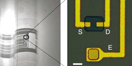
Left: optical micrograph of ECoG probe conforming onto a curvilinear surface (scale bar, 1 mm); right: optical micrograph of the channel of a transistor and a surface electrode, in which the gold films act as source (S), drain (D) and electrode pad (E) (scale bar, 10mm.) (credit: Department of Bioelectronics, Ecole des Mines)
“To understand how the brain works, we record the activity of a large number of neurons. Transistors provide higher-quality recordings than electrodes — and, in turn, record more neuronal activity,” said George Malliaras of the Microelectronics Center of Provence, France, and a lead author on the research. “The CNF prototyping allowed us to skip having to reinvent the wheel and saved us precious time and money.”

Layouts of the surface gold electrode (E, left) and of the transistor channel (right), showing transistor source (S) and drain (D), parylene insulator film, and PEDOT:PSS conductive polymer (credit: Department of Bioelectronics, Ecole des Mines)
The researchers fabricated highly conformable organic electrochemical transistors* (OECTs) and used them to measures ECoG signals on the somatosensory cortex of rats.
The study, “In Vivo Recordings of Brain Activity Using Organic Transistors,” was published in Nature Communications (open access) March 2013.
To develop the prototypes, the scientists used Cornell’s CNF lithography and characterization suite of tools.
The System Neuroscience Institute, Marseille, France, and Microvitae Technologies, Gardanne, France, also contributed to the research. The CNF facility is funded by the National Science Foundation.
* In contrast to FETs, where an oxide separates the channel from the electrolyte and prohibits any ion transport between these two layers, the channel of OECTs is in direct
contact with an electrolyte. As a result, the channel/electrolyte interface constitutes an integral part of the operation mechanism of OECTs. State-of-the-art OECTs are based on the conducting polymer poly(3,4-ethylenedioxythiophene) doped with polystyrene
sulfonate (PEDOT:PSS).