Quantum-assisted nano-imaging
May 6, 2013

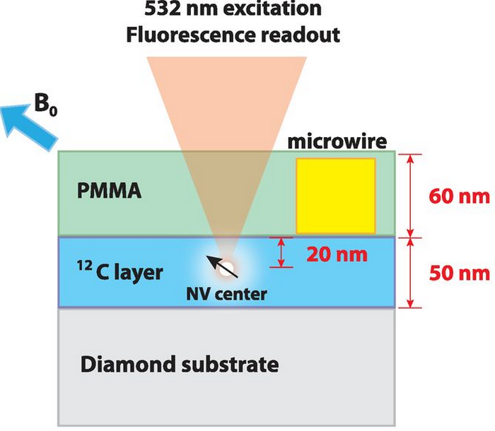
Basic configuration of NV-NMR detection, showing sample geometry along the diamond axis with NV spin embedded 20-nm deep within 12C diamond layer. The NV center detects NMR of protons in the PMMA polymer layer. (Credit: H.J. Mamin et al./Science)
Two separate teams of researchers working on DARPA’s Quantum-Assisted Sensing and Readout (QuASAR) program, led by the University of Stuttgart in Germany and IBM’s Almaden Research Center, have developed a nanoscale magnetometer that enables magnetic resonance imaging (MRI) with sufficient resolution to measure as few as 10,000 protons in a volume of only 125 cubic nanometers, which approaches the level of individual protein molecules.
Previous MRI technologies, even when stretched to their limits, have not allowed for resolution beyond a few microns because of distorting factors like background magnetic noise. The QuASAR teams’ new “nanoMRI” technique overcomes this limitation by using a single nitrogen-vacancy (NV) color center embedded close to the surface of a diamond chip to measure nuclear magnetic resonance signals.
It can be used to measure the magnetic field at a single point on a structure, or scan across the surface to image the structure by measuring multiple points. The nanoMRI technology offers the additional benefit of working at room temperatures, eliminating the need for expensive cryogenic equipment. Traditional MRI uses bulky machinery that frequently requires cryogenic cooling.

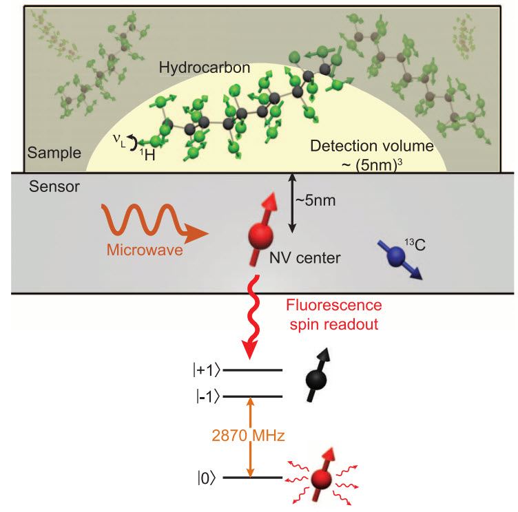
NV centers implanted near the diamond surface were used to detect proton spins within liquid and solid organic samples placed on the crystal surface (credit: T. Staudacher et al./Science)
The University of Stuttgart and IBM’s work on QuASAR could potentially offer a range of future medical benefits and capabilities:
- Support future drug development by facilitating increased understanding of the structure of proteins.
- Enable detailed, three-dimensional mapping of biological molecules, with sufficient sensitivity to identify specific elements. This information could streamline assessment of inhibitor drugs against naturally occurring and bioengineered viruses.
- Enable measurement of the magnetic field of firing neurons.
Future work may include attempts to increase the sensitivity of the measurement devices by moving them even closer to the organisms to be measured, embed nano-diamonds with NV centers into living cells for in-vitro studies to measure magnetic fields and temperature, and enable NV-assisted magnetic-field imaging of tagged bio-molecules.
The QuASAR nanoMRI technique was also used in a study (previously reported on KurzweilAI) by Harvard-Smithsonian Center for Astrophysics (CfA) scientists in developing a method for determining the magnetic structure of living “magnetotactic” bacteria (MTB) down to a sub-cellular level.