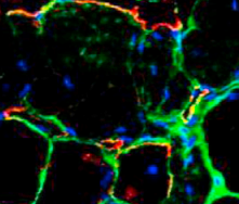
Researchers grow human muscle cells in a dish
November 12, 2013
Skeletal muscle has proved to be very difficult to grow in patients with muscular dystrophy and other disorders that degrade and weaken muscle. But researchers at Boston Children’s Hospital‘s Stem Cell Program now report boosting muscle mass and reversing disease in a mouse model of Duchenne muscular dystrophy, using a “cocktail” of three compounds identified through a new rapid culture system.
Adding the same compounds to stem cells derived from patients’ skin cells, they then successfully grew human muscle cells in a dish.
Senior investigator Leonard Zon, MD, director of the Stem Cell Program at Boston Children’s and an investigator with the Howard Hughes Medical Institute, hopes for clinical trials to begin transplanting these cells into patients with muscle loss in the next several years. In the meantime, the screening technique, described in the journal Cell (open access), can also be applied to multiple disorders beyond muscular dystrophy, he says.
The researchers found that a combination of three small molecules (forskolin, basic fibroblast growth factor and a glycogen synthase kinase-3 GSK-3beta inhibitor) was able to reprogram iPS cells into muscle cells that successfully engrafted in mice.
Zon’s next goal is to transplant these muscle cells from the dish into patients through clinical trials he hopes develop within the next several years. He may begin by trying to increase muscle mass in a specific anatomic location, but for a condition like Duchenne muscular dystrophy, he envisions a series of local injections in different parts of the body.
Zon believes that the combination of rapid zebrafish screening with patient-specific iPS cells will greatly speed up therapeutic development, allowing scientists to quickly vet large numbers of compounds and possible culture conditions. They can then enter a much shorter list of compounds into lengthier testing in human iPS cells that recapitulate the disease in question, as well as live mouse models.
The zebrafish system can also quickly spot inhibitors of tissue growth; in fact, it grew out of the lab’s desire to find chemicals that blocked muscle development to treat rhabdomyosarcoma, a tumor that grows in skeletal muscle. The Zon Lab is also using it to find chemicals that can block expansion of melanocytes, potentially halting melanoma.
Using these technologies, the Zon Lab has developed and filed a patent on a platform for large-scale chemical screening in zebrafish. The lab has also benefited from funding from the Massachusetts Life Sciences Center.
Abstract of Cell paper
Ex vivo expansion of satellite cells and directed differentiation of pluripotent cells to mature skeletal muscle have proved difficult challenges for regenerative biology. Using a zebrafish embryo culture system with reporters of early and late skeletal muscle differentiation, we examined the influence of 2,400 chemicals on myogenesis and identified six that expanded muscle progenitors, including three GSK3β inhibitors, two calpain inhibitors, and one adenylyl cyclase activator, forskolin. Forskolin also enhanced proliferation of mouse satellite cells in culture and maintained their ability to engraft muscle in vivo. A combination of bFGF, forskolin, and the GSK3β inhibitor BIO induced skeletal muscle differentiation in human induced pluripotent stem cells (iPSCs) and produced engraftable myogenic progenitors that contributed to muscle repair in vivo. In summary, these studies reveal functionally conserved pathways regulating myogenesis across species and identify chemical compounds that expand mouse satellite cells and differentiate human iPSCs into engraftable muscle.
