Rice University installs powerful electron microscope with sub-nanoscale resolution
June 30, 2015

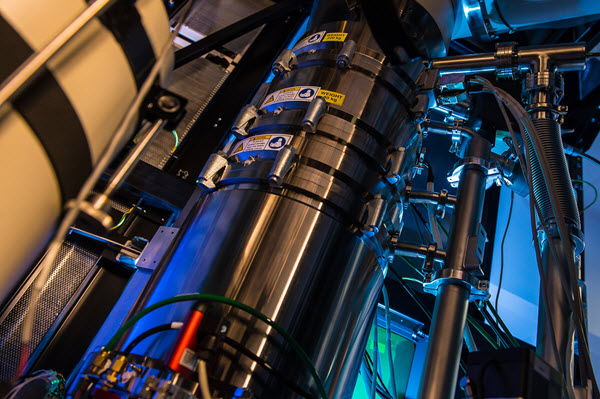
The Titan Themis microscope at Rice University incorporates a variety of detectors, including X-ray, optical, and multiple electron detectors and a 4K-resolution camera. The microscope gives researchers the ability to create three-dimensional structural reconstructions and carry out electric field mapping of subnanoscale materials. (credit: Jeff Fitlow/Rice University)
Rice University has installed the Titan Themis scanning/transmission electron microscope, which will enable scientists from Rice as well as academic and industrial partners to view and analyze materials at angstrom-scale (one-tenth of a nanometer) resolution, about the size of a single hydrogen atom.
Images will be captured with a variety of detectors, including X-ray, optical and multiple electron detectors and a 4K-resolution camera (will create 4K ultra HD images). The microscope gives researchers the ability to create three-dimensional structural reconstructions and carry out electric field mapping of subnanoscale materials.
Rice University | The Titan microscope gives Rice researchers a new look at atoms
Electron microscopes use beams of electrons rather than light to illuminate objects of interest. Because the wavelength of electrons is so much smaller than that of photons, the microscopes are able to capture images of much smaller things with greater detail than even the highest-resolution optical microscope. Titan is a fourth-generation model manufactured in the Netherlands. It’s the latest and most powerful model and the first to be installed in the United States.
“The beauty of these newer instruments is their analytical capabilities,” said said Emilie Ringe, a Rice assistant professor of materials science and nanoengineering and of chemistry. “Now we can probe a particular atom’s chemical composition. Through various techniques, either via scattering intensity, X-rays emission or electron-beam absorption, we can figure out, say, that we’re looking at a palladium atom or a carbon atom. We couldn’t do that before.”
Another instrument, a Helios NanoLab 600 DualBeam microscope, will be used for three-dimensional imaging, analysis of larger samples, and preparation of thin slices of samples for the more powerful Titan next door.
“A visual image of something on an atomic level can give you so much more information than a few numbers can,” said Peter Rossky, a theoretical chemist and dean of Rice’s Wiess School of Natural Sciences. Comparing images of the same material taken by an older electron microscope and the Titan Themis was like “the difference between a black-and-white TV and high-definition color,” he said.
“Taking a complex image — not just a picture but a spectrum image that has lots of energy information — in the older model would take about 35 minutes,” she said. “By that time, the electron beam has destroyed whatever you were trying to look at. With this generation, you have the data you need in about two minutes. You can generate a lot more data more quickly.”
Rice plans to host a two-day workshop in September to introduce the microscopes and their capabilities to the research community at the university and beyond. Beginning this summer, Ringe said, the electron microscopy center will be open to Rice students and faculty as well as researchers from other universities and industry.