Scientists create first 3-D synchronized-beating heart tissue
February 8, 2017

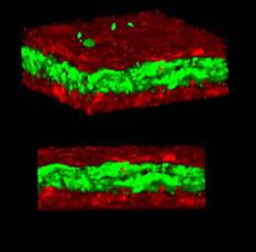
3-D fluorescent image of 3-D tissue with many cells laid down sequentially to create attached layers of alternating cell types, like membranes in the human body (credit: York University)
York University scientists have created the first in vitro (lab) 3D heart tissue made from three different types of cardiac cells that beat in synchronized harmony. It may lead to better understanding of cardiac health and improved treatments.*
The researchers constructed the heart tissue from three free-beating rat cell types: contractile cardiac muscle cells, connective tissue cells, and vascular cells. No external scaffold was used and the cells were the only building blocks of the generated cardiac tissue. The researchers believe this is the first 3D in vitro cardiac tissue with three cell types that can beat together as one entity, rather than at different intervals, with high cell density and efficient cell contacts, and without the requirement of external electrical stimulation.
The substance used to stick the cells together (ViaGlue) may also provide researchers with tools to create and test 3D in vitro cardiac tissue in their own labs to study heart disease and issues with transplantation.
“This breakthrough will allow better and earlier drug testing, and potentially eliminate harmful or toxic medications sooner,” said York U chemistry Professor Muhammad Yousaf.
For 2D or 3D cardiac tissue to be functional it needs the same high cellular density and the cells must be in contact to facilitate synchronized beating, according to the researchers. The 3D cardiac tissue was created at a millimeter scale, but larger versions could be made, said Yousaf, who has created a start-up company, OrganoLinX, to commercialize the ViaGlue reagent and to provide custom 3D tissues on demand.
“Production of 3-dimensional artificial cardiac tissues for fundamental studies of heart disease, transplantation, and evaluation of drug toxicity is an important and intense area of research,” the researchers note in a paper in open-access Nature Scientific Reports.
* Cardiovascular-associated diseases are the leading cause of death globally and are responsible for 40 per cent of deaths in North America, according to a 2011 report from the American Heart Association.
York University | York U makes a 3D heart beat as one
Abstract of Scaffold Free Bio-orthogonal Assembly of 3-Dimensional Cardiac Tissue via Cell Surface Engineering
There has been tremendous interest in constructing in vitro cardiac tissue for a range of fundamental studies of cardiac development and disease and as a commercial system to evaluate therapeutic drug discovery prioritization and toxicity. Although there has been progress towards studying 2-dimensional cardiac function in vitro, there remain challenging obstacles to generate rapid and efficient scaffold-free 3-dimensional multiple cell type co-culture cardiac tissue models. Herein, we develop a programmed rapid self-assembly strategy to induce specific and stable cell-cell contacts among multiple cell types found in heart tissue to generate 3D tissues through cell-surface engineering based on liposome delivery and fusion to display bio-orthogonal functional groups from cell membranes. We generate, for the first time, a scaffold free and stable self assembled 3 cell line co-culture 3D cardiac tissue model by assembling cardiomyocytes, endothelial cells and cardiac fibroblast cells via a rapid inter-cell click ligation process. We compare and analyze the function of the 3D cardiac tissue chips with 2D co-culture monolayers by assessing cardiac specific markers, electromechanical cell coupling, beating rates and evaluating drug toxicity.