Tracing the brain’s connections to dopamine neurons
June 15, 2012

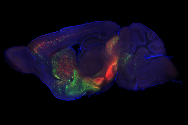
Direct inputs to dopamine neurons: green dots represent neurons providing inputs to dopamine neurons in the ventral tegmental area (VTA), while red dots represent neurons providing inputs to SNc dopamine neurons (credit: Mitsuko Watabe-Uchida and Sachie Ogawa)
A genetically modified version of the rabies virus is helping scientists at Harvard create the first comprehensive list of inputs that connect directly to dopamine neurons, a research effort that could help lead to treatments for Parkinson’s disease and addiction.
Dopamine plays a major role in the brain system that is responsible for reward-driven learning.
The researchers, led by Naoshige Uchida, associate professor of molecular and cellular biology, used the virus in two parts of the brain: the ventral tegmental area (VTA), known for processing reward, and the substantia nigra (SNc), known for motor control, to find other parts of the brain that connect directly to dopamine neurons.
“By understanding their inputs, we might be able to better understand how the function of dopamine neurons is regulated, and, in turn, how addiction happens, and how Parkinson’s disease and other motor-control disorders are affected by problems with dopamine neurons,” Uchida said.
“And because this application provides us with very quantitative data, it’s possible that this is a technique that might be useful in attacking the causes of those diseases.”
Both the VTA and SNc have high concentrations of dopamine neurons, but Uchida also chose to examine both areas because the cells in the two regions fire differently.
“We wanted to know what the difference was, generally,” Uchida said. “That’s why we compared the inputs to both structures. Based on how other neurons are connected there, we can start to explain why these two regions of the brain do different things.”
How to visually trace neural connections
The challenge, however, is that dopamine neurons are packed into relatively small regions with several other cell types. To ensure they were only observing dopamine neurons, researchers turned to an organism more typically known for damaging neurons: the rabies virus.
Before the researchers infect genetically engineered mice with the rabies virus, however, they first injected the animals with a pair of “helper” viruses. The first causes dopamine neurons to produce a receptor protein, meaning that the rabies virus can only infect dopamine neurons, while the second restores the virus’ ability to “hop” from one neuron to another.
The mice are then infected with a version of the rabies virus that has been genetically modified to produce a fluorescent protein, allowing researchers to track the virus as it binds with dopamine neurons, and then jumps to the cells that link directly to those neurons.
The results, as seen in images of a mouse’s brain depicting the wealth of connections to dopamine neurons, show that a number of brain regions — including some previously unknown areas — are connected to dopamine neurons.
“We found some new connections, and we found some that we suspected were there, but that were not well understood,” Uchida said. “For example, we found that there are connections between the motor cortex and the SNc, which may be related to SNc dopamine neurons’ role in motor control.
Deep brain stimulation clue
“Other connections, though, were more intriguing,” he continued. “We found that the subthalamic nucleus preferentially connects to SNc neurons. That’s particularly important because that region is a popular target for deep brain stimulation as a treatment for Parkinson’s.”
“The mechanism for why deep brain stimulation works is not completely understood,” Uchida said. “There was speculation that it might have been inhibiting neurons in the subthalamic nucleus. But our findings suggest, because there is a direct connection between those neurons and dopamine neurons in the SNc, that it is actually activating those neurons. I don’t think this explains the entire mechanism for why deep brain stimulation works, but this may be part of it.
“This work also offers us a roadmap for other areas we might investigate, so now we can target those areas and record from them,” Uchida added. “This is a critical step for future investigations.”
Funding for the research was provided by the Howard Hughes Medical Institute, the Richard and Susan Smith Family Foundation, the Alfred P. Sloan Foundation, and the Milton Fund.
Ref.: Mitsuko Watabe-Uchida, Lisa Zhu, Sachie K. Ogawa, Archana Vamanrao, Naoshige Uchida, Whole-Brain Mapping of Direct Inputs to Midbrain Dopamine Neurons, Neuron, 2012, DOI: 10.1016/j.neuron.2012.03.017