Ultrasound treats Alzheimer’s disease, restoring memory in mice
March 17, 2015

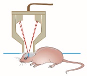
Scanning ultrasound treatment of Alzheimer’s disease in mouse model (credit: Gerhard Leinenga and Jürgen Götz/Science Translational Medicine)
University of Queensland researchers have discovered that non-invasive scanning ultrasound (SUS) technology* can be used to treat Alzheimer’s disease in mice and restore memory by breaking apart the neurotoxic Amyloid-β (Aβ) peptide plaques that result in memory loss and cognitive decline.
The method can temporarily open the blood-brain barrier (BBB), activating microglial cells that digest and remove the amyloid plaques that destroy brain synapses.
Treated AD mice displayed improved performance on three memory tasks: the Y-maze, the novel object recognition test, and the active place avoidance task.
The next step is to scale the research in higher animal models ahead of human clinical trials, which are at least two years away. In their paper in the journal Science Translational Medicine, the researchers note possible hurdles. For example, the human brain is much larger, and it’s also thicker than that of a mouse, which may require stronger energy that could cause tissue damage. And it will be necessary to avoid excessive immune activation.
The researchers also plan to see whether this method clears toxic protein aggregates in other neurodegenerative diseases and restores executive functions, including decision-making and motor control. It could also be used as a vehicle for drug or gene delivery, since the BBB remains the major obstacle for the uptake by brain tissue of therapeutic agents.
Previous research in treating Alzheimer’s with ultrasound used magnetic resonance imaging (MRI) to focus the ultrasonic energy to open the BBB for more effective delivery of drugs to the brain.

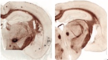
Scanning ultrasound plaque reduction in mouse-model hippocampus. (Left) without treatment. (Right) with treatment. Compact mature plaques are shown in amber and more diffuse amyloid deposits are shown in black. Quantification of amyloid plaques revealed a 56% reduction in the area of cortex occupied by plaques. (credit: Gerhard Leinenga and Jürgen Götz/Science Translational Medicine)
* The researchers used 0.7-MPa peak rarefactional pressure, 10-Hz pulse repetition frequency, 10% duty cycle, 1 MHz center frequency, and 6-s sonication time per spot.
Abstract of Scanning ultrasound removes amyloid-β and restores memory in an Alzheimer’s disease mouse model
Amyloid-β (Aβ) peptide has been implicated in the pathogenesis of Alzheimer’s disease (AD). We present a nonpharmacological approach for removing Aβ and restoring memory function in a mouse model of AD in which Aβ is deposited in the brain. We used repeated scanning ultrasound (SUS) treatments of the mouse brain to remove Aβ, without the need for any additional therapeutic agent such as anti-Aβ antibody. Spinning disk confocal microscopy and high-resolution three-dimensional reconstruction revealed extensive internalization of Aβ into the lysosomes of activated microglia in mouse brains subjected to SUS, with no concomitant increase observed in the number of microglia. Plaque burden was reduced in SUS-treated AD mice compared to sham-treated animals, and cleared plaques were observed in 75% of SUS-treated mice. Treated AD mice also displayed improved performance on three memory tasks: the Y-maze, the novel object recognition test, and the active place avoidance task. Our findings suggest that repeated SUS is useful for removing Aβ in the mouse brain without causing overt damage, and should be explored further as a noninvasive method with therapeutic potential in AD.