Uncovering the spread of deadly cancer
August 30, 2011

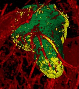
Image of Glioblastoma multiforme, a brain cancer, obtained using a new imaging technique. The main tumor is green, blood vessels feeding the tumor are red, and migrating cells, yellow. (Credit: Case Western Reserve University School of Medicine)
Researchers at Case Western Reserve University School of Medicine have imaged individual cancer cells and the routes they travel as the tumor spreads, allowing the scientists to see pathways to stop a deadly brain cancer.
Current molecular imaging techniques suffer from low resolution and difficulty in imaging through the skull.
The researchers used a mouse model that included four different cell lines of brain cancers at various stages of tumor development and dispersion. The cancer cells were modified with fluorescent markers and implanted in the model’s brain.
To get a look at glioblastoma multiforme, a particularly aggressive cancer that has no known treatments to stop it from spreading, the researchers used a novel cryo-imaging (cold temperature) technique. Using custom algorithms, the cryo-imaging system disassembled the brain, layer by layer, and reassembled it into a color three-dimensional digital image.
The researchers were able to differentiate the main tumor mass, the blood vessels that feed the cancer, and dispersing cells. The new imaging system enabled them to peer at single cells and analyze the extent and patterns of cancer cell migration and dispersal from tumors along blood vessels and white matter tracts within the brain, offering exquisite anatomic detail.
They found that two cell lines, a human brain cancer LN229, and a rodent cancer CNS-1, best resemble the actions of glioblastoma multiforme in human patients. This novel cryo-imaging technique provides a valuable tool to evaluate therapeutic interventions targeted at limiting tumor cell invasion and dispersal, they concluded.
Ref.: S. M. Burden-Gulley, et al., Novel Cryo-Imaging of the Glioma Tumor Microenvironment Reveals Migration and Dispersal Pathways in Vivid Three-Dimensional Detail, Cancer Research, 2011; [DOI: 10.1158/0008-5472.CAN-11-1553]