Visualizing how TMS affects groups of neurons in real time
September 17, 2014

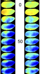
Spatiotemporal activity patterns induced by a single TMS pulse (left) and 10 Hz TMS (right) over 50 milliseconds (credit: Vladislav Kozyre et al./PNAS)
German neuroscientists have developed a method for recording the effects of transcranial magnetic stimulation (TMS) on animals in real time, using voltage-sensitive dyes, which emit fluorescent light signals that indicate which groups of neurons are activated or inhibited.
Using fMRI is too slow to show real-time effects, and with rapid measurement methods like EEG and MEG, the TMS magnetic field generates artifacts.
So RUB researchers headed by Dirk Jancke, PhD, of the Institut für Neuroinformatik (Ruhr-University Bochum), used voltage-sensitive dyes, which emit fluorescent light showing changes in the cells’ membrane potential across several square millimeters of cortical tissue when neurons are activated or inhibited, and can respond in microseconds.
“We can now demonstrate in real time how a single TMS pulse suppresses brain activity across a considerable region, most likely through mass activation of inhibiting brain cells,” said Jancke.
The German Research Foundation funded the study.
Abstract of Proceedings of the National Academy of Sciences paper
Transcranial magnetic stimulation (TMS) is widely used in clinical interventions and basic neuroscience. Additionally, it has become a powerful tool to drive plastic changes in neuronal networks. However, highly resolved recordings of the immediate TMS effects have remained scarce, because existing recording techniques are limited in spatial or temporal resolution or are interfered with by the strong TMS-induced electric field. To circumvent these constraints, we performed optical imaging with voltage-sensitive dye (VSD) in an animal experimental setting using anaesthetized cats. The dye signals reflect gradual changes in the cells’ membrane potential across several square millimeters of cortical tissue, thus enabling direct visualization of TMS-induced neuronal population dynamics. After application of a single TMS pulse across visual cortex, brief focal activation was immediately followed by synchronous suppression of a large pool of neurons. With consecutive magnetic pulses (10 Hz), widespread activity within this “basin of suppression” increased stepwise to suprathreshold levels and spontaneous activity was enhanced. Visual stimulation after repetitive TMS revealed long-term potentiation of evoked activity. Furthermore, loss of the “deceleration–acceleration” notch during the rising phase of the response, as a signature of fast intracortical inhibition detectable with VSD imaging, indicated weakened inhibition as an important driving force of increasing cortical excitability. In summary, our data show that high-frequency TMS changes the balance between excitation and inhibition in favor of an excitatory cortical state. VSD imaging may thus be a promising technique to trace TMS-induced changes in excitability and resulting plastic processes across cortical maps with high spatial and temporal resolutions.