Worm ‘EEG’ tests neural effects of drugs
May 24, 2013

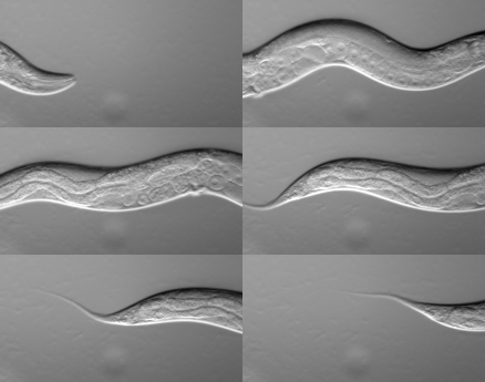
C. elegans movement (credit: Wikimedia Commons)
Scientists from the University of Southampton have developed a microfluidic electrophysiological device called a NeuroChip that records the neural activity in the microscopic worm Caenorhadbitis elegans (C. elegans) — the worm equivalent of an EEG —.to help test the effects of drugs.
How to record a worm’s ‘EEG’
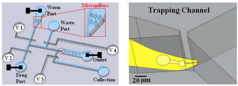
With the NeuroChip, you feed the worm into a narrow, fluid-filled channel that tapers at one end. That captures the worm by its “head,” so now it’s now immobilized — the correct orientation for recording the activity of the nervous system. The metal electrodes are connected to an amplifier to make the recording.
C. elegans is a nematode (roundworm) that has been enormously important in providing insight into fundamental signaling processes in the nervous system. It’s small (about 1 mm in length), transparent for ease of manipulation and observation, feeds on bacteria such as E. coli, can be easily and cheaply housed and cultivated in large numbers (10,000 worms/petri dish) in the laboratory, and has a short life cycle (speeds up testing).
However, electrophysiological recordings of the activity of excitatory and inhibitory nerve cells in the nervous system of the worm previously required a high level of technical expertise. Single microscopic (1mm long) worms had to be carefully trapped on the end of a glass tube (microelectrode) to make the recording. The worms are very mobile and tiny, so this can be a challenging procedure.
As the worm turns

Diagram of the NeuroChip (left). It is a two-layer structure: the blue layer is the microfluidic region, processing worms and delivery of drugs. Trapping region (red circled) is magnified (right), showing the trapped worm’s head. The red square is the micro-pillar region, which facilitates correct orientation of the worm. The white layer is the pneumatic control layer (V1, V2, V3 and V4 indicate Valve 1, 2, 3 and 4 respectively). The three black squares are the microelectrodes. (Credit: Chunxiao Hu et al./PLoS ONE)
The new design simplifies the process, making it more efficient, and improves the signal to noise (quality) of the recorded data.
The NeuroChip can detect the effects of drugs and allows for high-throughput screens; researchers can quickly conduct millions of chemical, genetic or pharmacological tests in neurotoxicology and generic screening for neuroactive drugs, the researchers say.
According to Lindy Holden-Dye, Professor of Neuroscience at the University of Southampton and lead author of the paper, “We are particularly interested in using this as a sensitive new tool for screening compounds for neurotoxicity. It will allow us to precisely quantify sub-lethal effects on neural network activity. It can also provide an information-rich platform by reporting the effects of compounds on a diverse array of neurotransmitter pathways, which are implicated in mammalian toxicology. ”
The research is a joint project between the University’s Centre for Biological Sciences and the Hybrid Biodevices Group.