stories on progressBreakthrough 3D images show how newborn head deforms in childbirth
January 1, 2020
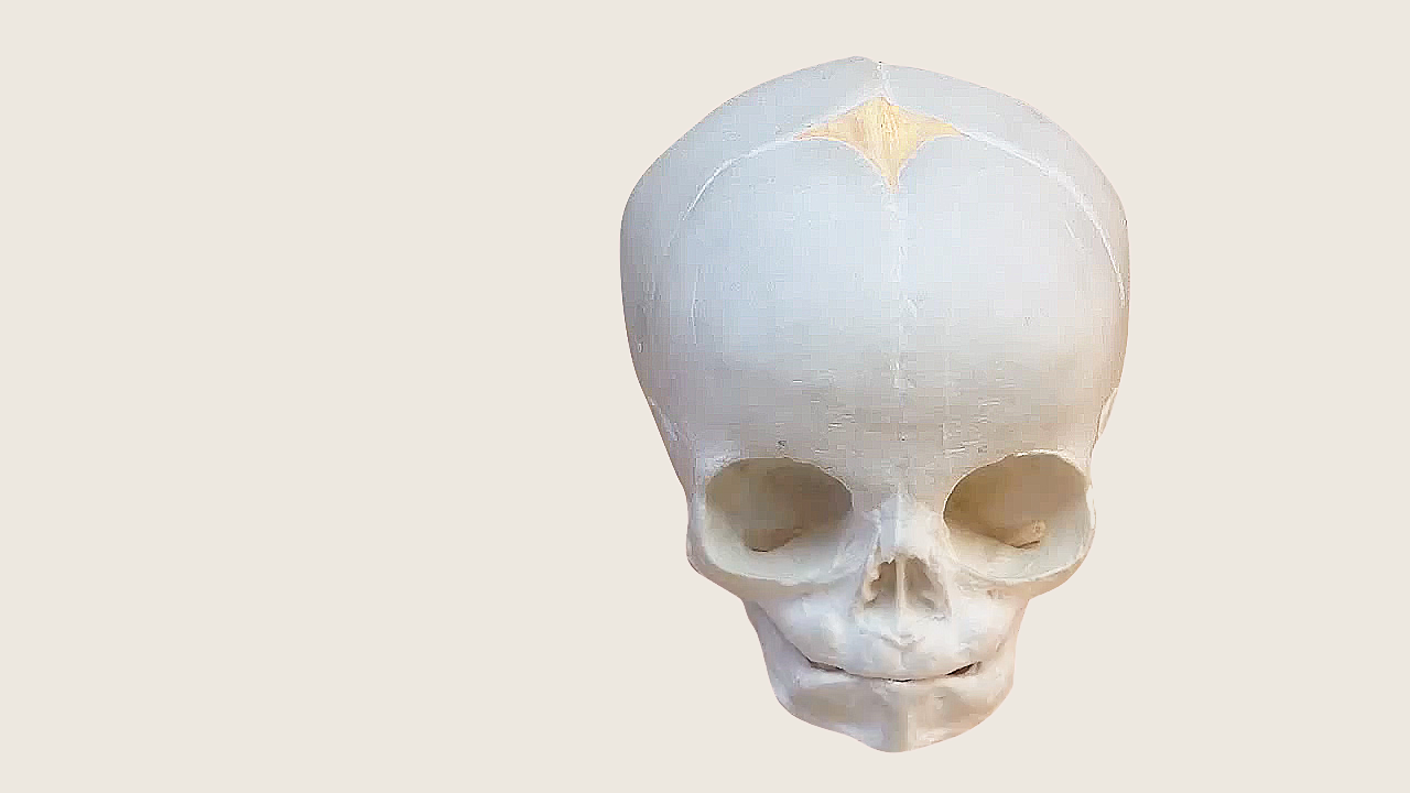
image | above
Model of an infant skull showing its separated bone plates.
— contents —
~ story
~ gallery
~ featurette
~ sketches
~ reading
— story —
An introduction.
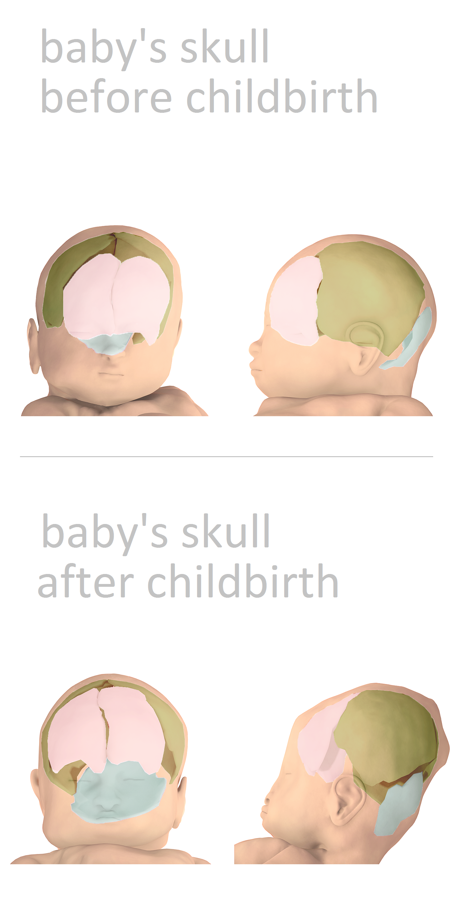
When babies pass through the mother’s birth canal during childbirth, the tight fit temporarily squashes their tiny heads — elongating their flexible skulls. The pressure dramatically changes the shape of their brain. For a new study, French data scientists + medical researchers have created 3D images that show the extensive cone-shaped distortion. The study was conducted in association with the university hospital center of the Univ. Clermont Auvergne in France.
A baby’s head can naturally change shape under pressure, because the bones in infant skulls haven’t fused together yet. There are soft areas at the top of the baby’s head to cope with being squeezed through the birth canal — so the newborn can be pushed-out with less head trauma.
Then the soft regions begin to harden and fuse over time. This makes room for the brain to grow during infancy, but protects it from impact as the child starts moving around. It can take 9 to 18 months before a baby’s skull is fully formed.
But the precise mechanics of how a baby’s skull + brain change shape during labor are not well understood. To learn more about that process — and how it can lead to injury — the French scientists conducted magnetic resonance imaging (MRI) scans of 7 pregnant women:
- when they were between weeks 36 –to– 39 of their pregnancies ( third trimester )
- and again when they were in labor, after her cervix was fully dilated
The problem of newborn head molding.
Their images revealed significant skull squeezing — known as newborn (or fetal) head molding —– in all the infants, and showed that the mechanical pressures on infant heads + brains during childbirth are stronger than we once thought.
In all 7 fetuses, skull bones that did not overlap prior to labor: were visibly overlapped once labor began, deforming the newborn’s head and brain. In 5 babies, the skulls returned to their pre-labor shape soon after childbirth — and the deformity was not noticeable when the newborns were examined.
Some valuable insights.
The MRI scans successfully captured views of soft tissues that were not visible with ultra-sound imaging. These visuals gave the physicians important clues to understand:
- deformation of fetal skulls + brains
- movement of the mother’s soft tissues around the baby during childbirth
The researchers said these results could be used someday to help create a ‘virtual labor’ — like a walk-through model showing the probable risks of your baby’s birth, before it happens. That could give mothers and their obstetric doctors early warning, so they can plan how to move forward with the childbirth.
image gallery | below
The 3D digital reconstruction — from MRI scans — of the infants’ brain + skull bones:
- in the womb before labor
- and during the 2nd stage of labor = when the infant is being pushed through the birth canal.
HALFLINE
presented by
PLoS One | home ~
tag line: t
the Public Library of Science | home ~
tag line: t
BABY no. 1 |
The 3D reconstruction — from MRI scans — of the infant’s skull bones.
BABY no. 1 |
The 3D reconstruction — from MRI scans — of the infant’s brain.

BABY no. 2 |
The 3D reconstruction — from MRI scans — of the infant’s brain.

BABY no. 3 |
The 3D reconstruction — from MRI scans — of the infant’s brain.

Better understanding another head deformity illness.
The 3D models created by this study produced a deeper knowledge of newborn head molding than physicians had before. These results will help doctors better understand a similar childhood condition called deformational plagiocephaly. The name plagiocephaly means “sloping head” — from the Greek words plagio for sloping + cephale for head.
Children with deformational plagiocephaly have a dangerous injury. Their skull + brain became deformed from too much compression against a crib mattress — or another flat surface. This illness is accidentally caused by parents who don’t rotate their infant’s body when the baby is laying down for a long time, without moving.
If a baby doesn’t switch positions enough — the skull gets lots of continual pressure at the same spot on their head. Eventually the baby’s fluids and soft tissues move away from the pressure — flattening-out in one part of the head, and blobbing-out into another. That’s why the illness is nick-named “flat head” syndrome. It’s been linked to developmental disability.
To prevent this illness, doctors teach parents to rotate the baby’s body on a routine schedule, especially while sleeping. In severe cases, physicians customize a padded helmet for the child — to protect its head from pressure, and fix the deformation over time.
— featurette —
An anatomical tour of the infant skull.
IMAGE
images | below
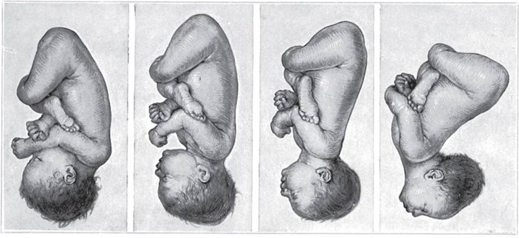
The pressures from the mother’s birth canal during childbirth cause a range of deformed head shapes called newborn (or fetal) head molding. These sketches from a medical book show a variety of common positions the baby can take while its exiting the womb — showing the way the head and brain deform in those postures.
book: Williams Obstetrics
year: 2018
publisher: McGraw Hill Education

The future of infant health.
It’s not well understood what the long-term prognosis is for a baby born with extreme newborn head molding. Even if the soft tissues and malleable bone eventually return to their normal shape — are there unseen + lasting consequences for that child’s health? Is there a risk of physical or cognitive disability? The startling images from the 3-D model leave parents with a lot of unanswered questions.
Does severe newborn head molding change the baby’s bone + tissues in a way that causes future risks: like micro-fractures, bone brittleness, vascular disorders, bruising problems? Or a diminished ability to heal from accidents or illness that affect the head?
Could babies born with extreme newborn head molding experience a higher rate of traumatic brain injury, later on? For example, from typical head impacts that happen to all kids — from falling down or playing sports? Will that child grow-up to have a lower endurance for brain concussion?
Can the common medical procedure called a Caesarean section (or C section) — where doctors surgically remove the infant through an abdominal incision— reduce fetal head molding that’s caused by squeezing through the birth canal?
A picture worth a thousand words.
In this study, physicians can clearly see the intense pressure on the baby’s head during childbirth. The good news is that researchers are finally getting the graphical evidence they need — so they can sort-out precisely what happens with newborn head molding.
The 3-D reconstruction paints a close-up picture of the baby’s experience. Data from the study showing: the shape, size, and timing of newborn head molding helps the medical team:
- build more accurate models
- make better predictions
- design preventative treatments
- develop medical interventions
- prepare for emergencies
Technology leads the way forward for medicine. Our health-care system is based entirely on what we can observe + understand. Modern tools — such as MRI and computer graphics — give us the ability to witness biology in motion.
image | below
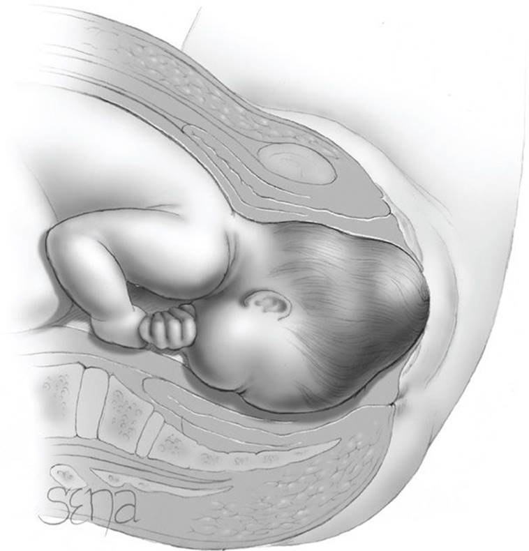
A sketch from a medical book showing a baby with the condition called newborn — or fetal — head molding.
HALFLINE
presented by
book: Williams Obstetrics
date: 2018
publisher: McGraw Hill Education | visit
IMAGE
reading
1. |
publication: Live Science
story title: Head deformity ignites debate among baby experts
deck: Has increased exponentially over 20 years.
read | story
HALFLINE
2. |
publication: Live Science
story title: How much do babies’ skulls get squished during birth?
deck: A whole lot, 3D images reveal
read | story
HALFLINE
presented by
text | home ~
tag line: text
3. |
publication: NewsWeek
story title: 3D MRI images show how a baby’s head shape changes during labor in incredible detail
read | story
HALFLINE
presented by
text | home ~
tag line: text
4. |
publication: One
story title: 3D MRI of fetal head molding + brain shape changes during 2nd stage of labor
read | story
HALFLINE
presented by
PLoS One | home
tag line:
— about —
PLoS One is a journal of the Public Library of Science.
PLoS | home
tag line:
BOTTOM
by definition | fetal ultra-sound
— notes —
MRI = magnetic resonance imaging
3D = 3-dimensional
PLoS = the Public Library of Science
CHU = university hospital center
