Engineers reveal fabrication process for revolutionary transparent graphene neural sensors
October 14, 2016

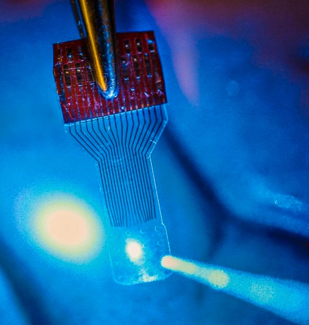
A blue light shines through a transparent, implantable medical sensor onto a brain. The invention may help neural researchers better view brain activity. (credit: Justin Williams research group)
In an open-access paper published Thursday (Oct. 13, 2016) in the journal Nature Protocols, University of Wisconsin–Madison engineers have published details of how to fabricate and use neural microelectrocorticography (μECoG) arrays made with transparent graphene in applications in electrophysiology, fluorescent microscopy, optical coherence tomography, and optogenetics.
Graphene is one of the most promising candidates for transparent neural electrodes, because the material has a UV to IR transparency of more than 90%, in addition to its high electrical and thermal conductivity, flexibility, and biocompatibility, the researchers note in the paper. That allows for simultaneous high-resolution imaging and optogenetic control.

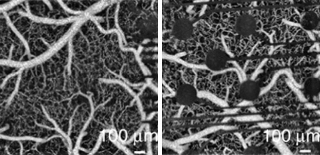
Left: Optical coherence tomography (OCT) image captured through an implanted transparent graphene electrode array, allowing for simultaneous observation of cells immediately beneath electrode sites during optical or electrical stimulation. Right: Optical coherence tomography (OCT) image taken with an implanted conventional opaque platinum electrode array. (credit: Dong-Wook Park et al./Nature Protocols)
The procedures in the paper are for a graphene μECoG electrode array implanted on the surface of the cerebral cortex and can be completed within 3–4 weeks by an experienced graduate student, according to the researchers. But this protocol “may be amenable to fabrication and testing of a multitude of other electrode arrays used in biological research, such as penetrating neural electrode arrays to study deep brain, nerve cuffs that are used to interface with the peripheral nervous system (PNS), or devices that interface with the muscular system,” according to the paper.
The researchers first announced the breakthrough in the open-access journal Nature Communications in 2014, as KurzweilAI reported. Now, the UW–Madison researchers are looking at ways to improve and build upon the technology. They also are seeking to expand its applications from neuroscience into areas such as research of stroke, epilepsy, Parkinson’s disease, cardiac conditions, and many others. And they hope other researchers do the same.
Funding for the initial research came from the Reliable Neural-Interface Technology program at the U.S. Defense Advanced Research Projects Agency.
The research was led by Zhenqiang (Jack) Ma, the Lynn H. Matthias Professor and Vilas Distinguished Achievement Professor in electrical and computer engineering at UW–Madison and Justin Williams, the Vilas Distinguished Achievement Professor in biomedical engineering and neurological surgery at UW–Madison.
Researchers at the University of Wisconsin-Milwaukee, Medtronic PLC Neuromodulation, the University of Washington, and Mahidol University in Bangkok, Thailand were also involved.
Abstract of Fabrication and utility of a transparent graphene neural electrode array for electrophysiology, in vivo imaging, and optogenetics
Transparent graphene-based neural electrode arrays provide unique opportunities for simultaneous investigation of electrophysiology, various neural imaging modalities, and optogenetics. Graphene electrodes have previously demonstrated greater broad-wavelength transmittance (~90%) than other transparent materials such as indium tin oxide (~80%) and ultrathin metals (~60%). This protocol describes how to fabricate and implant a graphene-based microelectrocorticography (μECoG) electrode array and subsequently use this alongside electrophysiology, fluorescence microscopy, optical coherence tomography (OCT), and optogenetics. Further applications, such as transparent penetrating electrode arrays, multi-electrode electroretinography, and electromyography, are also viable with this technology. The procedures described herein, from the material characterization methods to the optogenetic experiments, can be completed within 3–4 weeks by an experienced graduate student. These protocols should help to expand the boundaries of neurophysiological experimentation, enabling analytical methods that were previously unachievable using opaque metal–based electrode arrays.