First real-time imaging of neural activity invented
November 24, 2015

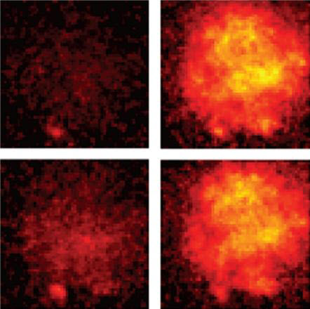
A series of images from a Duke engineering experiment show voltage spreading through a fruitfly neuron over a matter of just 4 milliseconds, a hundred times faster than the blink of an eye. The technology can see impulses as fleeting as 0.2 millisecond — 2000 times faster than a blink. (credit: Yiyang Gong, Duke University)
Researchers at Stanford University and Duke University have developed a new technique for watching the brain’s neurons in action with a temporal (time) resolution of about 0.2 milliseconds — a speed that is just fast enough to capture the action potentials in mammalian brains in real time for the first time.
The researchers combined genetically encoded voltage indicators, which can sense individual action potentials from many cell types in live animals, with a protein that can quickly sense neural voltage potentials with another protein that can amplify its signal output — the brightest fluorescing protein available.
The paper appeared online in Science on November 19, 2015.
Abstract of High-speed recording of neural spikes in awake mice and flies with a fluorescent voltage sensor
Genetically encoded voltage indicators (GEVIs) are a promising technology for fluorescence readout of millisecond-scale neuronal dynamics. Prior GEVIs had insufficient signaling speed and dynamic range to resolve action potentials in live animals. We coupled fast voltage-sensing domains from a rhodopsin protein to bright fluorophores via resonance energy transfer. The resulting GEVIs are sufficiently bright and fast to report neuronal action potentials and membrane voltage dynamics in awake mice and flies, resolving fast spike trains with 0.2-millisecond timing precision at spike detection error rates orders of magnitude better than prior GEVIs. In vivo imaging revealed sensory-evoked responses, including somatic spiking, dendritic dynamics, and intracellular voltage propagation. These results empower in vivo optical studies of neuronal electrophysiology and coding and motivate further advancements in high-speed microscopy.