First US patients treated with noninvasive focused ultrasound for Parkinson’s disease
September 2, 2015

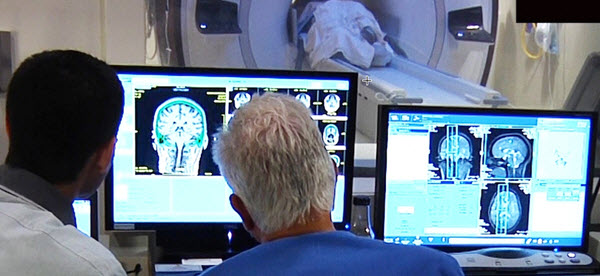
University of Maryland medical doctors monitor focused ultrasound treatment for essential tremor, guided by magnetic resonance imaging (MRI) (credit: University of Maryland School of Medicine)
Researchers at the University of Maryland have performed the first focused ultrasound treatments on a deep structure within the brain related to Parkinson’s disease* called the globus pallidus.
These treatments are part of international pilot studies of 40 patients assessing the feasibility, safety, and preliminary efficacy of focused ultrasound treatments for Parkinson’s disease, guided by magnetic resonance imaging (MRI).
The researchers are using MRI to help them guide ultrasound waves through the intact skin and skull to reach the globus pallidus part of the brain. If successful, focused ultrasound could offer an alternative approach for certain patients with Parkinson’s disease who have failed medical therapy or become disabled from medication-induced dyskinesia (tremor). To date, seven patients in Korea and one patient in Canada have been treated in studies.
The new Parkinson’s procedure

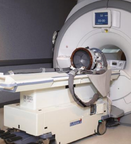
ExAblate Neuro system (credit: Insightec)
The non-invasive ultrasound and MRI imaging procedures are done on an outpatient basis in the Center for Metabolic Imaging and Image-Guided Therapeutics (CMIT) MRI suite, using the ExAblate Neuro system developed by Insightec.
During the Parkinson’s procedure, patients lie in an MRI scanner with a head-immobilizing frame fitted with a transducer helmet. Ultrasonic energy is targeted through the skull to the globus pallidus of the brain, and images acquired during the procedure give physicians a real-time map of the area being treated.
“We’re raising the temperature in a very restricted area of the brain to destroy tissue,” explained principal investigator Howard M. Eisenberg, MD, the Raymond K. Thompson Chair of Neurosurgery. “The ultrasound waves create a heat lesion that we can monitor through MRI.”
The entire procedure lasts two to four hours, and patients are awake and able to interact with the treatment team. This allows the physicians to monitor the immediate effects of treatment and make adjustments if necessary.
Researchers from the University of Virginia Health System reported in the New England Journal of Medicine in 2013 that 15 patients with essential tremor** — a related disorder — who received focused ultrasound saw “significant improvement” in their dominant hand tremor. Patients treated in the initial phase of the study at the University of Maryland experienced similar results.
The Michael J. Fox Foundation for Parkinson’s Research and the Focused Ultrasound Foundation are funding the new Parkinson’s study.
* As many as one million Americans have Parkinson’s disease, a chronic, degenerative disorder for which there is no cure. The second most common movement disorder, Parkinson’s results from the malfunction or loss of brain cells crucial for movement and coordination. Symptoms include motor difficulties such as tremor, rigidity and postural instability. People with Parkinson’s can also experience non-motor symptoms of cognitive impairment, depression, and anxiety, and autonomic dysfunction.
** Essential tremor, which is eight times more common than Parkinson’s disease, causes debilitating shaking that can be resistant to drug therapy. It mainly affects the hands, head and voice, making aspects of daily life like eating, drinking and writing extremely difficult.
Abstract of A Pilot Study of Focused Ultrasound Thalamotomy for Essential Tremor
Background
Recent advances have enabled delivery of high-intensity focused ultrasound through the intact human cranium with magnetic resonance imaging (MRI) guidance. This preliminary study investigates the use of transcranial MRI-guided focused ultrasound thalamotomy for the treatment of essential tremor.
Methods
From February 2011 through December 2011, in an open-label, uncontrolled study, we used transcranial MRI-guided focused ultrasound to target the unilateral ventral intermediate nucleus of the thalamus in 15 patients with severe, medication-refractory essential tremor. We recorded all safety data and measured the effectiveness of tremor suppression using the Clinical Rating Scale for Tremor to calculate the total score (ranging from 0 to 160), hand subscore (primary outcome, ranging from 0 to 32), and disability subscore (ranging from 0 to 32), with higher scores indicating worse tremor. We assessed the patients’ perceptions of treatment efficacy with the Quality of Life in Essential Tremor Questionnaire (ranging from 0 to 100%, with higher scores indicating greater perceived disability).
Results
Thermal ablation of the thalamic target occurred in all patients. Adverse effects of the procedure included transient sensory, cerebellar, motor, and speech abnormalities, with persistent paresthesias in four patients. Scores for hand tremor improved from 20.4 at baseline to 5.2 at 12 months (P=0.001). Total tremor scores improved from 54.9 to 24.3 (P=0.001). Disability scores improved from 18.2 to 2.8 (P=0.001). Quality-of-life scores improved from 37% to 11% (P=0.001).
Conclusions
In this pilot study, essential tremor improved in 15 patients treated with MRI-guided focused ultrasound thalamotomy. Large, randomized, controlled trials will be required to assess the procedure’s efficacy and safety. (Funded by the Focused Ultrasound Surgery Foundation; ClinicalTrials.gov number, NCT01304758.)