New microscope produces 3-D movies of live cells
March 6, 2011
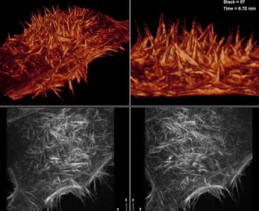
High-speed imaging reveals the surface of a HeLa cell (credit: Janelia Farm)
Scientists at the Howard Hughes Medical Institute Janelia Farm Research Campus have created a microscope that lets researchers see the dynamic inner lives of living cells using a form of high speed imaging called Bessel beam plane illumination microscopy.
The microscope uses an exquisitely thin sheet of light— similar to that used in supermarket bar-code scanners — to peer inside single living cells and create dazzling 3-D movies that make biological processes, such as cell division, come alive.
Their work appears March 4 in Nature Methods.
Janelia Farm group leader Eric Betzig comments that until recently, microscopes could see objects no smaller than 200 nanometers in size. Several years ago, Betzig and his Janelia Farm colleague Harald Hess invented photoactivated localization microscopy, PALM, which can produce images of objects only 10-20 nanometers in size.
Although other researchers, including Janelia Farm Fellow Philipp Keller, have used plane illumination to great effect to study multicellular organisms hundreds of microns in size, the light sheets were still too thick to work effectively for imaging within single cells only tens of microns in size.
The new microscope is also exciting because it may be used in the future to improve super-resolution microscopy. PALM and other super-resolution techniques are limited to looking at thin, dead samples, and can be very damaging when looking at live ones. Bessel beam plane illumination microscopy will be a powerful tool for cell biologists, Betzig says, since it noninvasively images the rapidly evolving three-dimensional complexity of cells.
Adapted from materials provided by Howard Hughes Medical Institute