Scientists Create 3D Models of Whole Mouse Organs
June 25, 2010
Yale University engineers have for the first time created 3D models of whole intact mouse organs.
Combining an imaging technique called multiphoton microscopy (using light to excite naturally fluorescent cells within the tissue) with “optical clearing” (using a solution that renders tissue transparent), the researchers were able to scan mouse organs and create high-resolution images of the brain, small intestine, large intestine, kidney, lung and testicles.

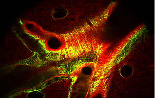
Collagen fibers (in green) outline the bronchiole pathways against a background of elastin tissue (in red) in this high-resolution image of a mouse lung. (Michael Leven/Yale)
They then created 3D models of the complete organs—a feat that, until now, was only possible by slicing the organs into thin sections for staining, destroying them in the process.
The new technique could be used to create 3D models of biopsies, said Michael Levene, associate professor at the Yale School of Engineering & Applied Science and the team leader. This could be especially useful in tissues where the direction of a cancerous growth may make it difficult to know how to slice tissue sample, he noted.
In addition, the technology could eventually be used to trace fluorescent proteins in the mouse brain and see where different genes are expressed, or to trace where drugs travel in the body using fluorescent tagging, for example.
More info: Yale University news