Ultrasound applied to brain found to elevate mood
July 22, 2013

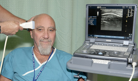
Transcranial ultrasound is applied with gel on the scalp overlying the frontal temporal cortex. Dr. Hameroff, shown here, tested the system on himself before conducting a study with patient volunteers. (Credit: The University of Arizona Health Sciences Center)
University of Arizona researchers have found in a recent study that ultrasound waves applied to specific areas of the brain appear able to alter patients’ moods.
The technique could one day be used to treat conditions such as depression and anxiety.
With research committee and hospital approval, and patient informed consent, Dr. Stuart Hameroff, professor emeritus of the UA’s departments of anesthesiology and psychology and director of the UA’s Center for Consciousness Studies, and colleagues applied transcranial ultrasound stimulation (TUS) to 31 chronic pain patients at The University of Arizona Medical Center-South Campus.
This was a double-blind study (neither doctor nor subject knew if the ultrasound machine had been switched on or off).

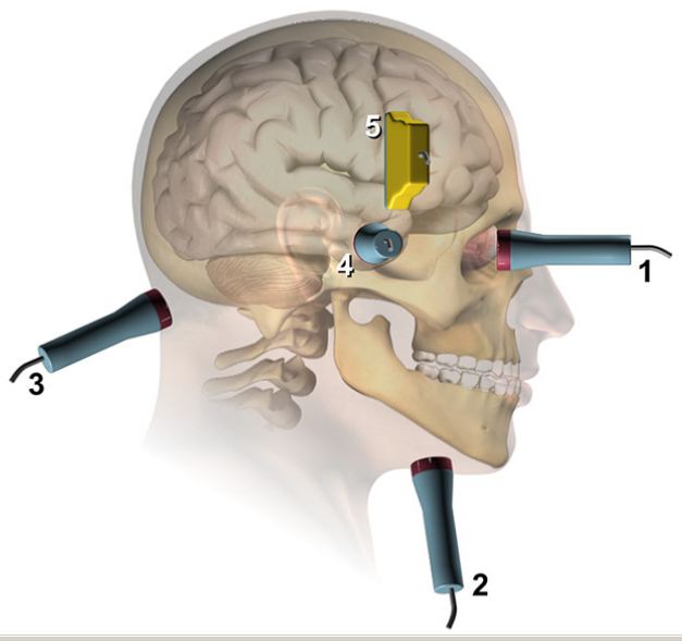
Sites for transcranial ultrasound (TUS). 1-4 are used in Doppler blood fl ow studies. 1: trans-orbital, 2: sub-mandibular, 3: sub-occipital, 4: temporal window. 5 is the site used in present study, overlying frontal-temporal cortex. (Credit: Stuart Hameroff et al./Brain Stimulation)
Patients reported improvements in mood for up to 40 minutes following treatment with brain ultrasound, compared with no difference in mood when the machine was switched off.
The researchers confirmed the patients’ subjective reports of increases in positive mood with a Visual Analog Mood Scale, or VAMS, a standardized objective mood scale often used in psychological studies.
“This was a pilot study which showed safety, and some efficacy, for clinical use of TUS, and help mediate mood and consciousness,” Hameroff said. TUS may benefit a variety of neurological and psychiatric disorders.”
The discovery may open the door to a possible range of new applications of ultrasound in medicine.
“We frequently use ultrasound in the operating room for imaging,” said Hameroff. “It’s safe as long as you avoid excessive exposure and heating.”
The mechanical waves, harmless at low intensities, penetrate the body’s tissues and bones, and an echo effect is used to generate images of anatomical structures such as fetuses in the womb, organs, and blood vessels.
Ultrasound may also be more desirable than existing brain stimulation techniques such as transcranial magnetic stimulation, or TMS. Used to treat clinically depressed patients, TMS can have side effects, including what some describe as an unpleasant sensation of magnetic waves moving through the head.

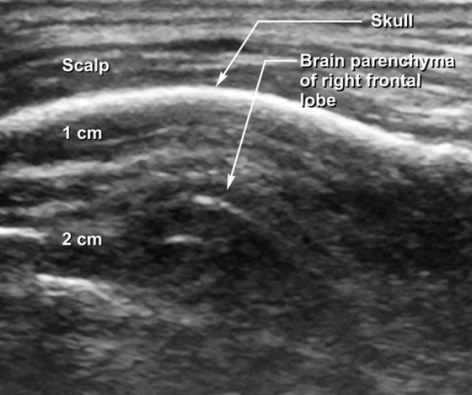
Transcranial ultrasound image
A study of TUS effects on mood
After finding promising preliminary results in chronic pain patients, Hameroff and colleagues set out to discover whether transcranial ultrasound stimulation could improve mood in a larger group of healthy volunteer test subjects.
Jay Sanguinetti, a doctoral candidate in the department of psychology and his adviser John Allen, a UA distinguished professor of psychology, conducted a followup study of ultrasound on UA psychology student volunteers.
They recorded vital signs such as heart rate and breath rate, and narrowed down the optimum treatment to 2 megahertz for 30 seconds as the most likely to produce a positive mood change in patients.
“With 2 megahertz, those who were stimulated with ultrasound reported feeling ‘lighter,’ or ‘happier;’ a little more attentive, a little more focused, and a general increase in well-being,” Sanguinetti said.

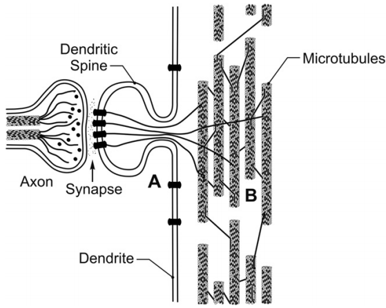
Two theories of how ultrasound works. William J. Tyler, School of Life Sciences, Arizona State University proposed that ultrasound affected viscoelastic properties of neuronal membranes and extracellular fluid, altering membrane conductance (zones marked by A). Hameroff and colleagues suggest that ultrasound acts via microtubules (B), which have resonant frequencies in the megahertz range. Here, axon terminal releases neurotransmitters into synapse and onto receptors in membrane of dendritic spine. Actin filaments (as well as soluble second messengers, not shown) connect to cytoskeletal microtubules in main dendrite. Two microtubules are seen in the axon (left); dendritic microtubules (right) are arranged in local networks, interconnected by microtubule-associated proteins (MAPs). (Artwork credit: Dave Cantrell)
Allen and Sanguinetti then began a double-blind clinical trial to verify the statistical significance of their findings and to rule out any possibility of a placebo effect in their patients. Results of the trials are being analyzed, Sanguinetti said.
“What we think is happening is that the ultrasound is making the neurons a little bit more likely to fire in the parts of the brain involved with mood,” thus stimulating the brain’s electrical activity and possibly leading to a change in how participants feel, Sanguinetti said.
Wearable ultrasound device
The UA researchers are collaborating with Silicon Valley-based Neurotrek, which is developing a device that potentially could target specific regions of the brain with ultrasound.
The UA researchers will work with a prototype of the Neurotrek device to test its efficacy and potential applications.
“The idea is that this device will be a wearable unit that noninvasively and safely interfaces with your brain using ultrasound to regulate neural activity,” said Sanguinetti.