‘Wearable’ PET brain scanner enables studies of moving patients
May 23, 2017

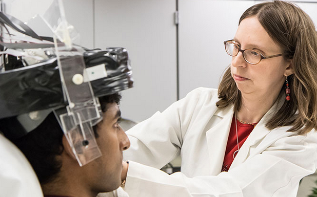
Julie Brefczynski-Lewis, a neuroscientist at West Virginia University, places a helmet-like PET scanner on a research subject. The mobile scanner enables studies of human interaction, movement disorders, and more. (credit: West Virginia University)
Two scientists have developed a miniaturized positron emission tomography (PET) brain scanner that can be “worn” like a helmet.
The new Ambulatory Microdose Positron Emission Tomography (AMPET) scanner allows research subjects to stand and move around as the device scans, instead of having to lie completely still and be administered anesthesia — making it impossible to find associations between movement and brain activity.

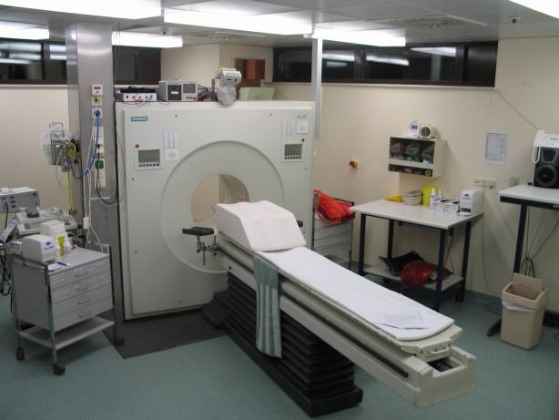
Conventional positron emission tomography (PET) scanners immobilize patients (credit: Jens Maus/CC)
The AMPET scanner was developed by Julie Brefczynski-Lewis, a neuroscientist at West Virginia University (WVU), and Stan Majewski, a physicist at WVU and now at the University of Virginia. It could make possible new psychological and clinical studies on how the brain functions when affected by diseases from epilepsy to addiction, and during ordinary and dysfunctional social interactions.

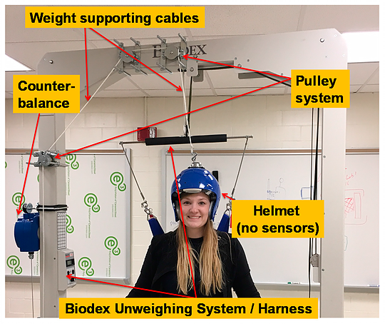
Helmet support prototype with weighted helmet, allowing for freedom of movement. The counterbalance currently supports up to 10 kg but can be upgraded. Digitizing electronics will be mounted to the support above the patient. (credit: Samantha Melroy et al./Sensors)
Because AMPET sits so close to the brain, it can also “catch” more of the photons stemming from the radiotracers used in PET than larger scanners can. That means researchers can administer a lower dose of radioactive material and still get a good biological snapshot. Catching more signals also allows AMPET to create higher resolution images than regular PET.

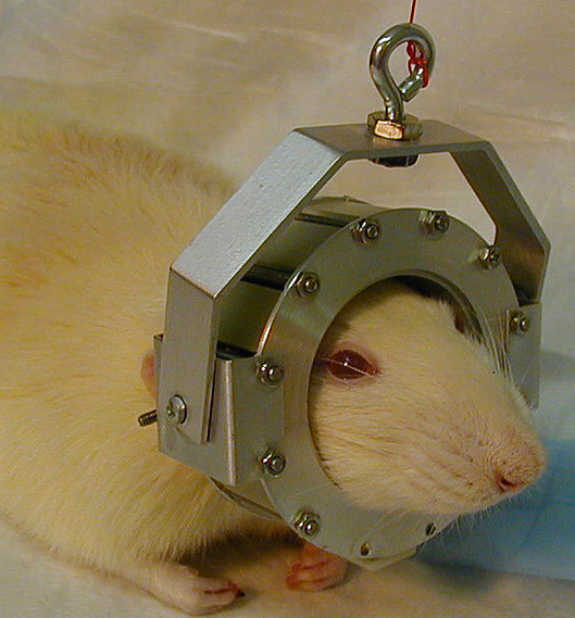
The AMPET idea was sparked by the Rat Conscious Animal PET (RatCAP) scanner for studying rats at the U.S. Department of Energy’s (DOE) Brookhaven National Laboratory.** The scanner is a 250-gram ring that fits around the head of a rat, suspended by springs to support its weight and let the rat scurry about as the device scans. (credit: Brookhaven Lab)
The researchers plan to build a laboratory-ready version next.
Seeing more deeply into the brain

A patient or animal about to undergo a PET scan is injected with a low dose of a radiotracer — a radioactive form of a molecule that is regularly used in the body. These molecules emit anti-matter particles called positrons, which then manage to only travel a tiny distance through the body. As soon as one of these positrons meets an electron in biological tissue, the pair annihilates and converts their mass to energy. This energy takes the form of two high-energy light rays, called gamma photons, that shoot off in opposite directions. PET machines detect these photons and track their paths backward to their point of origin — the tracer molecule. By measuring levels of the tracer, for instance, doctors can map areas of high metabolic activity. Mapping of different tracers provides insight into different aspects of a patient’s health. (credit: Brookhaven Lab)
PET scans allow researchers to see farther into the body than other imaging tools. This lets AMPET reach deep neural structures while the research subjects are upright and moving. “A lot of the important things that are going on with emotion, memory, and behavior are way deep in the center of the brain: the basal ganglia, hippocampus, amygdala,” Brefczynski-Lewis notes.
“Currently we are doing tests to validate the use of virtual reality environments in future experiments,” she said. In this virtual reality, volunteers would read from a script designed to make the subject angry, for example, as his or her brain is scanned.
In the medical sphere, the scanning helmet could help explain what happens during drug treatments. Or it could shed light on movement disorders such as epilepsy, and watch what happens in the brain during a seizure; or study the sub-population of Parkinson’s patients who have great difficulty walking, but can ride a bicycle .
The RatCAP project at Brookhaven was funded by the DOE Office of Science. RHIC is a DOE Office of Science User Facility for nuclear physics research. Brookhaven Lab physicists use technology similar to PET scanners at the Relativistic Heavy Ion Collider (RHIC), where they must track the particles that fly out of near-light speed collisions of charged nuclei. PET research at the Lab dates back to the early 1960s and includes the creation of the first single-plane scanner as well as various tracer molecules.
Abstract of Development and Design of Next-Generation Head-Mounted Ambulatory Microdose Positron-Emission Tomography (AM-PET) System
Several applications exist for a whole brain positron-emission tomography (PET) brain imager designed as a portable unit that can be worn on a patient’s head. Enabled by improvements in detector technology, a lightweight, high performance device would allow PET brain imaging in different environments and during behavioral tasks. Such a wearable system that allows the subjects to move their heads and walk—the Ambulatory Microdose PET (AM-PET)—is currently under development. This imager will be helpful for testing subjects performing selected activities such as gestures, virtual reality activities and walking. The need for this type of lightweight mobile device has led to the construction of a proof of concept portable head-worn unit that uses twelve silicon photomultiplier (SiPM) PET module sensors built into a small ring which fits around the head. This paper is focused on the engineering design of mechanical support aspects of the AM-PET project, both of the current device as well as of the coming next-generation devices. The goal of this work is to optimize design of the scanner and its mechanics to improve comfort for the subject by reducing the effect of weight, and to enable diversification of its applications amongst different research activities.