Cutting-edge MRI techniques for studying communication within the brain
February 9, 2012

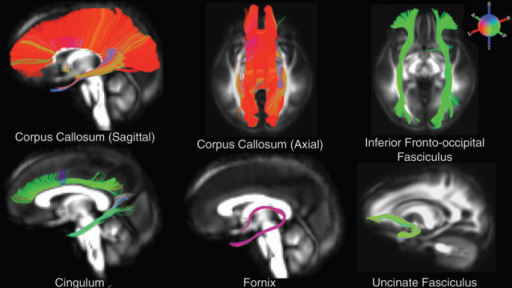
White matter (WM) tract reconstructions for several major WM pathways.(credit: Andrew L. Alexander et al./Brain Connectivity)
Researchers from the University of Wisconsin, Madison have presented innovative magnetic resonance imaging (MRI) techniques that can measure changes in the microstructure of the white matter likely to affect brain function and the ability of different regions of the brain to communicate.
Brain function depends on the ability of different brain regions to communicate through signaling networks that travel along white matter tracts.
Using different types and amounts of tissue staining to measure how water molecules interact with the surrounding brain tissue, researchers can quantify changes in the density, orientation, and organization of white matter. They can then use this information to generate image maps of these signaling networks, a method called tractography.
Andrew Alexander and colleagues from University of Wisconsin, Madison describe three quantitative MRI (qMRI) techniques that are enabling the characterization of the microstructural properties of white matter: diffusion MRI, magnetization transfer imaging, and relaxometry. This approach can be used to study and compare the properties of brain tissue across populations and to shed light on mechanisms underlying aging, disease, and gender differences in brain function, for example.
“White matter is the material that provides for the wiring and connectivity between brain regions. This exciting paper describes three new methodologies to measure the integrity of white matter in normal and diseased brain. These methods show promise in multiple sclerosis, depression, aging, and human development,” says Bharat Biswal, PhD, Co-Editor-in-Chief of Brain Connectivity and Associate Professor, University of Medicine and Dentistry of New Jersey.
Ref.: Andrew L. Alexander, et al., Characterization of Cerebral White Matter Properties Using Quantitative Magnetic Resonance Imaging Stains, Brain Connectivity, 2012; [DOI:10.1089/brain.2011.0071]