Mapping connectomes in the visual cortex
April 11, 2011

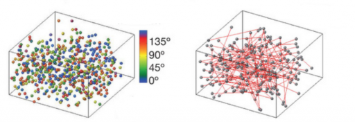
Visual neurons responding to various visual-edge angles (left) and connections for the 135 degrees angle (right) (H. Ko et al./Nature)
Scientists at University College London have developed a technique that enables them to combine information about the function of neurons together with details of their synaptic connections, finding that connection probability is related to the similarity of visually driven neuronal activity.
The researchers used high-resolution imaging of neurons in the visual cortex of mouse brains, which contains thousands of neurons and millions of different connections. Using high-resolution imaging, they were able to detect which of these neurons responded to a particular stimulus, for example, a horizontal edge.
Taking a slice of the same tissue, the researchers then applied small currents to a subset of neurons in turn to see which other neurons responded and which of them were synaptically connected. By repeating this technique many times, the researchers were able to trace the function and connectivity of hundreds of nerve cells in the visual cortex.
Resolving a longstanding debate, the researchers showed that neurons that responded similarly to visual stimuli, such as those that respond to edges of the same orientation, tend to connect to each other much more than those that prefer different orientations.
Ref.: H. Ko et al., Functional specificity of local synaptic connections in neocortical networks, April 10 online edition, Nature