Sharper, deeper, faster optical imaging of live biological samples
July 26, 2011

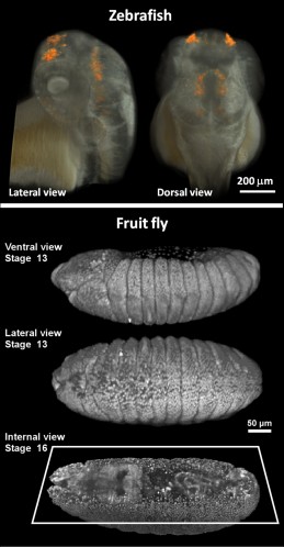
Three-dimensional live imaging of zebrafish (upper panel) and fruit fly (lower panel) embryos with two-photon light sheet microscopy (credit: Willy Supatto, Seth Ruffins, Thai Truong/Caltech)
Researchers from the California Institute of Technology (Caltech) have developed a novel approach that could redefine optical imaging of live biological samples, simultaneously achieving high resolution, high penetration depth (for seeing deep inside 3D samples), and high imaging speed.
The research team employed an unconventional imaging method called light-sheet microscopy: a thin, flat sheet of light is used to illuminate a biological sample from the side, creating a single illuminated optical section through the sample.
The light given off by the sample is then captured with a camera oriented perpendicularly to the light sheet, harvesting data from the entire illuminated plane at once. This allows millions of image pixels to be captured simultaneously, reducing the light intensity that needs to be used for each pixel.
To achieve sharper image resolution with light-sheet microscopy deep inside biological samples, the team used a process called two-photon excitation for the illumination.
“The goal is to create ‘digital embryos,’ providing insights into how embryos are built, which is critical not only for basic understanding of how biology works but also for future medical applications such as robotic surgery, tissue engineering, or stem-cell therapy,” said biologist Scott Fraser, director of the Biological Imaging Center at Caltech’s Beckman Institute.
Thai V. Truong, et al., Deep and fast live imaging with two-photon scanned light-sheet microscopy, Nature Methods, 2011; [DOI: 10.1038/nmeth.1652]