Stanford researcher scans his own brain for a year and a half — the most studied in the world
December 16, 2015

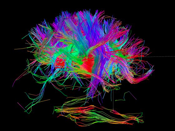
Human connectome (Credit: NIH Human Connectome Project)
You’ve probably seen the “connectome” map of the major networks between different functional areas of the human brain. Cool graphic. But this is just an average.
It raises a lot of questions: How does this map relate to your brain? Do these connections persist over a period of months or more? Or do they vary with different conditions (happy or sad mood, etc.)? And what if you’re a schizophrenic, alcoholic, meditator, or videogamer, etc., how does your connectome look?
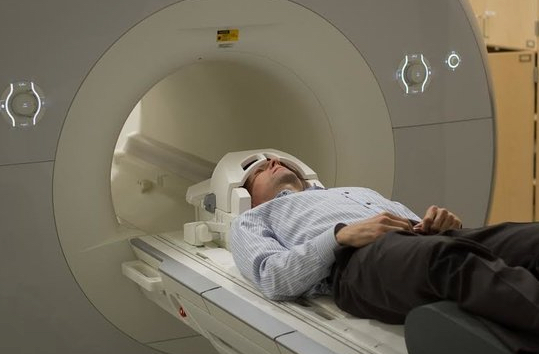
These questions obsessed Stanford psychologist Russell Poldrack, leading to his “MyConnectome project.” In the noble DIY tradition of Marie Curie, Jonas Salk, and Albert Hoffman, he started off his day by climbing into an MRI machine and scanning his brain for 10 minutes Tuesdays and Thursdays every week for a year and a half — making his brain the most studied in the world.

Poldrack’s morning FMRI scan (credit: Russell Poldrack)
He also fasted and drew blood on Tuesdays for testing with metabolomics (chemical fingerprints in biological fluids) and genomics (gene tests, performed by 23andMe).
The results — the most complete study of the brain’s network connections over time — are published in open-access Nature Communications.

An overview of the resting-state fMRI analysis pipeline (credit: Russell A. Poldrack et al./Nature Communications)
Here is some of what he found out:
- His connectivity was surprisingly consistent, which is good news for researchers studying differences between healthy brains and those of patients with neurological disorders that might suffer from disrupted connectivity, such as schizophrenia or bipolar disorder.
- There was a strong correlation between brain activity and changes in the expression of many different families of genes. The expression of genes related to inflammation and immune response matched Poldrack’s psoriasis flare-ups, for example.
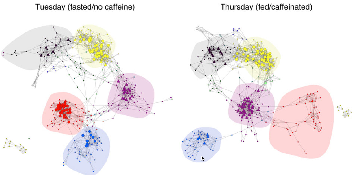
- Fasting with no caffeine on Tuesdays radically changed the connection between the somatosensory motor network and the higher vision network: it grew significantly tighter without caffeine. “That was totally unexpected, but it shows that being caffeinated radically changes the connectivity of your brain,” Poldrack said. “We don’t really know if it’s better or worse, but it’s interesting that these are relatively low-level areas. It may well be that I’m more fatigued on those days, and that drives the brain into this state that’s focused on integrating those basic processes more.”

Network connections for Tuesdays (fasted) and Thursdays (fed/caffeinated). Hubs are shown as larger nodes, with provincial hubs depicted as circles and connector hubs depicted as triangles. Network module membership is coded by node color; major networks are shaded, including somatomotor (red), second visual (blue), cingulo-opercular (purple), fronto-parietal (yellow) and default mode (black). (credit: Russell A. Poldrack et al./Nature Communications)
What’s next
“I’m generally a pretty happy and even-keeled person,” Poldrack said. “My positive mood is almost always high, and my negative mood is almost always non-existent. It would be interesting to scan people with a wider emotional variation and see how their connections look over time.” As he suggests in the video (below), “We need to learn a lot more about how individual brains differ from one another. … There are many more questions yet to be answered. … When it comes to understanding the brain. we really just scratched the surface.”
Fortunately, Poldrack and his colleagues have made the entire data set and the ready-built tools to analyze it available here. The data set is large and deep; Poldrack said he hopes people will approach it from innovative angles and uncover connections that will help advance the research. Meanwhile, Poldrack plans to hone software to elucidate the interplay between brain function and gene expression.
But so far we only have a experimental population of one. Any volunteers (and funders) for a follow-up study?
* In any action that a person undertakes, many different regions of the brain communicate with each other, serving as a sort of check-and-balance system to make sure that the correct actions are taken to deal with the situation at hand. These messages are communicated over more than a dozen networks, sets of functional areas of the brain that preferentially talk to one another.
There are multiple networks for vision, a somatosensory/motor network, and there are others that are attributed to attention or task management. Collectively, these are known as the connectome. Because the strength or efficiency of these individual networks can affect behavior, they have become of greater interest to researchers in recent years. To isolate these connections, researchers examine functional MRI data collected while the patient is at rest.
Stanford | Stanford researcher scans his own brain for a year and a half
Abstract of Long-term neural and physiological phenotyping of a single human
Psychiatric disorders are characterized by major fluctuations in psychological function over the course of weeks and months, but the dynamic characteristics of brain function over this timescale in healthy individuals are unknown. Here, as a proof of concept to address this question, we present the MyConnectome project. An intensive phenome-wide assessment of a single human was performed over a period of 18 months, including functional and structural brain connectivity using magnetic resonance imaging, psychological function and physical health, gene expression and metabolomics. A reproducible analysis workflow is provided, along with open access to the data and an online browser for results. We demonstrate dynamic changes in brain connectivity over the timescales of days to months, and relations between brain connectivity, gene expression and metabolites. This resource can serve as a testbed to study the joint dynamics of human brain and metabolic function over time, an approach that is critical for the development of precision medicine strategies for brain disorders.